Download
1 / 53
590 likes | 1.13k Views
Spine fracture. Anatomy of The Low Back. Spinal segment consists of the following: Two vertebrae An intervertebral disc between the two vertebrae Two nerve roots that leave the spinal cord, one on each side . Cont ….

E N D
Spinal segment consists of the following: Two vertebrae An intervertebral disc between the two vertebrae Two nerve roots that leave the spinal cord, one on each side Cont …
1. Protect the spinal cord, nerve roots, and internal organs.2. Provide flexibility of motion.3. Provide structural support and balance for upright posture. Spine bears the load of the head, shoulders and arms, and upper body. The upper body weight is then distributed to the hips and legs. The spine attempts to keep the body's weight balanced over the pelvis. This reduces the amount of work required by the spinal muscles and can eliminate muscle fatigue and back pain. Functions of the spine
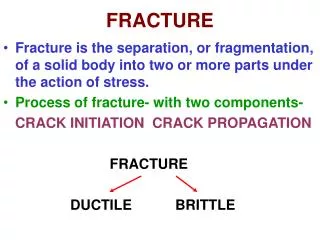
1. Anterior column consists of the anterior longitudinal ligament and the anterior part of the vertebral body. 2. Middle column extends from the middle portion of the vertebral body to the posterior aspect of the vertebral body and includes the posterior longitudinal ligament. 3. Posterior column includes all bony and ligamentous structures posterior to the posterior longitudinal ligament and includes the pedicles, facets, spinous processes and all associated ligaments. Fractures involving only the anterior columns are considered stable, while fractures that involve the middle or all three columns are considered unstable. Denis Classification: The Three-column Concept
Incidence of spine injury • Male : female = 4 : 1 • Injury is common at the cervicothoracic and throacolumbar regions Mode of injury : • RTA : 45 % • Falls : 20 % • Sports ( diving ) injuries : 15 % • Acts of violence : 15 %
Injuries of the cervical spine Common areas : • C1 – 2 and C5 – 7 ( common ) • Neurological damage ------ 40 % • Radiograph normal --------- 10 % Causes : • Fall from height • Diving injuries • RTA ( whiplash injury ) • Gunshot injuries
Mechanism of injury • Pure flexion force : fall from height • Flexion rotation force : fall on one side of the shoulder • Axial compression : fall of an object on the head results in load compression • Extension force : e.g ; whiplash injury • Lateral flexion • Direct injuries : assault , gunshot injury
Whiplash injury ( acceleration injury , cervical sprain syndrome , soft tissue neck injury ) • An injury caused by the neck and head being thrown suddenly backward then forward upon impact. • Impact forces the neck and head beyond their normal range of movement, causing tissue damage and pain.
Symptoms : - Headache Pain in the shoulders Pain between the shoulder blades Pain in one or both arms Rarely : vertigo , auditory or visual disturbance Neck pain/stiffness Low back pain Signs : - Loss of motion in the neck Neck muscle spasm Clinical features Investigation : - X – ray ---- usually normal MRI ------ to make diagnosis
Treatment Conservative : First aid • First 24 hours : - • an ice bag applied to the neck ( relieve inflammation) for 20 minutes at a time, Medication • nonsteroidal anti-inflammatory drugs, • antidepressants, • muscle relaxants, and • cervical collar (for 2 to 3 weeks). • Range of motion exercises, • physical therapy, and • cervical traction cervical collar
Exercise • Movement of deep muscles in the back of the head : Stand against a door or a wall with your head facing forward and move your eyes so you look towards the two, four, eight, and 10 o'clock positions. Repeat this a few times. • Bend the back of the head carefully forward as if taking a bow. Return to the starting position with your head straight and facing forward. • Draw the chin in towards your neck and bend the head carefully forward. Return to the starting position. Bend the head backwards far enough to look at the ceiling. Return to the starting position.
Cont.. • Tilt the head sideways, if possible try to maintain the glance at a fixed point at eye level. Return to the starting position. Repeat this action with the head tilted to the other side. • Turn the head as if trying to look backwards over the shoulder, first to the left and then to the right. Imagine following a horizontal line on the wall at eye level. • Place the ball between the wall and the forehead and then try to move it around on the wall in circles or figures of eights. Repeat the exercise, this time placing the ball between the back of the head and the wall.
Clinical features • History of trauma • Pain , • swelling and • inability to move neck • Tenderness and palpable gap over the involved spinous process • Neurological involvement • Injuries of the spinal cord at the cervical region
Ask the patient to raise both their arms in front of them simultaneously as strongly as then can while the examiner provides resistance to this movement. Compare the strength of each arm. The deltoid muscle is innervated by the C5 nerve root via the axillary nerve. Motor System ExaminationDeltoid muscle
Test the strength of lower arm flexion by holding the patient's wrist from above and instructing them to "flex their hand up to their shoulder". Provide resistance at the wrist. Repeat and compare to the opposite arm. This tests the biceps muscle. The biceps muscle is innervated by the C5 and C6 nerve roots via the musculocutaneous nerve. Biceps muscle
Ask the patient to extend their forearm against the examiner's resistance. Make certain that the patient begins their extension from a fully flexed position because this part of the movement is most sensitive to a loss in strength. This tests the triceps. Note any asymmetry in the other arm. The triceps muscle is innervated by the C6 and C7 nerve roots via the radial nerve. Triceps muscle
Test the strength of wrist extension by asking the patient to extend their wrist while the examiner resists the movement. This tests the forearm extensors. Repeat with the other arm. The wrist extensors are innervated by C6 and C7 nerve roots via the radial nerve. The radial nerve is the "great extensor" of the arm: it innervates all the extensor muscles in the upper and lower arm. Forearm extensors
Examine the patient's hands. Look for intrinsic hand, thenar and hypothenar muscle wasting. Test the patient's grip by having the patient hold the examiner's fingers in their fist tightly and instructing them not to let go while the examiner attempts to remove them. Normally the examiner cannot remove their fingers. This tests the forearm flexors and the intrinsic hand muscles. Compare the hands for strength asymmetry. Finger flexion is innervated by the C8 nerve root via the median nerve. Forearm flexors and the intrinsic hand muscles
Test the intrinsic hand muscles by having the patient abduct or "fan out" all of their fingers. Instruct the patient to not allow the examiner to compress them back in. Normally, one can resist the examiner from replacing the fingers. Finger abduction or "fanning" is innervated by the T1 nerve root via the ulnar nerve. Intrinsic hand muscles
Test the strength of the thumb opposition by telling the patient to touch the tip of their thumb to the tip of their pinky finger. Apply resistance to the thumb with your index finger. Repeat with the other thumb and compare. Thumb opposition is innervated by the C8 and T1 nerve roots via the median nerve. Thumb opposition
First test the flexion of the hip by asking the patient to lie down and raise each leg separately while the examiner resists. Repeat and compare with the other leg. This tests the iliopsoas muscles. Hip flexion is innervated by the L2 and L3 nerve roots via the femoral nerve. Iliopsoas muscles. Hip flexion
Test the adduction of the legs by placing your hands on the inner thighs of the patient and asking them to bring both legs together. This tests the adductors of the medial thigh. Adduction of the hip is mediated by the L2, L3 and L4 nerve roots. Adductors of the medial thigh
Test the abduction of the legs by placing your hands on the outer thighs and asking the patient to move their legs apart. This tests the gluteus maximus and gluteus minimus. Abduction of the hip is mediated by the L4, L5 and S1 nerve roots. Gluteus maximus and gluteus minimus.
Test the extension of the hip by instructing the patient to press down on the examiner's hand which is placed underneath the patient's thigh. Repeat and compare to the other leg. This tests the gluteus maximus. Hip extension is innervated by the L4 and L5 nerve roots via the gluteal nerve. Gluteus maximus.
Test extension at the knee by placing one hand under the knee and the other on top of the lower leg to provide resistance. Ask the patient to "kick out" or extend the lower leg at the knee. Repeat and compare to the other leg. This tests the quadriceps muscle. Knee extension by the quadriceps muscle is innervated by the L3 and L4 nerve roots via the femoral nerve. Quadriceps muscle
Test flexion at the knee by holding the knee from the side and applying resistance under the ankle and instructing the patient to pull the lower leg towards their buttock as hard as possible. Repeat with the other leg. This tests the hamstrings. The hamstrings are innervated by the L5 and S1 nerve roots via the sciatic nerve. Hamstrings
Test dorsiflexion of the ankle by holding the top of the ankle and have the patient pull their foot up towards their face as hard as possible. Repeat with the other foot. This tests the muscles in the anterior compartment of the lower leg. Ankle dorsiflexion is innervated by the L4 and L5 nerve roots via the peroneal nerve. Muscles of the anterior compartment
Holding the bottom of the foot, ask the patient to "press down on the gas pedal" as hard as possible. Repeat with the other foot and compare. This tests the gastrocnemius and soleus muscles in the posterior compartment of the lower leg. Ankle plantar flexion is innervated by the S1 and S2 nerve roots via the tibial nerve. Muscles in the posterior compartment
Ask the patient to move the large toe against the examiner's resistance "up towards the patient's face". The extensor halucis longus muscle is almost completely innervated by the L5 nerve root. This tests the extensor halucis longus muscle. Extensor halucis longus muscle.
Spinal cord is the largest nerve in the body. Nerves are cord-like structures made up of nerve fibers. Nerve fibers are responsible for the communication systems of the body, which include sensory, motor, and autonomic functions. The nerve fibers within the spinal cord carry messages between the brain and the rest of the body. Anatomy and Physiology
Nerves that lie within the spinal cord are upper motor neurons (UMNs). They carry the messages back and forth from the brain to the spinal nerves along the spinal tract. The spinal nerves that branch out from the spinal cord to the other parts of the body are lower motor neurons (LMNs). These spinal nerves exit and enter at each vertebral level and communicate with specific areas of the body. The sensory portion of the LMNs carry messages to the brain about sensation from the skin and other body parts and organs. The motor portion of the LMNs send messages from the brain to the various body parts to initiate actions such as muscle movement. Cont ..
The vertebral column, or spinal column, is made up of 4 regions : - Seven cervical vertebrae protect the eight cervical nerves; Twelve thoracic vertebrae protect the twelve thoracic nerves; Five lumbar vertebrae protect the five lumbar nerves; Five sacral vertebrae, which are fused as one bone, help protect the five sacral nerves Cont ..
As the body grows, the vertebral column grows more in length than the spinal cord, which usually ends between the first and second lumbar vertebrae. From this point the lumbar and sacral nerves branch out from the spinal cord and descend inside the spinal column before leaving the vertebral column at their corresponding vertebrae. Because of this fact there is a difference between the skeletal or bony level of vertebral fracture and the neurological level of spinal cord injury Cont …
What Happens after A Spinal Cord Injury? • Spinal cord injury (SCI) refers to any injury of the neural (pertaining to nerves) elements within the spinal canal. SCI can occur from either trauma or disease to the vertebral column or the spinal cord itself. • Most spinal cord injuries are the result of trauma to the vertebral column. Such trauma can cause a fracture of bone or tearing of ligaments with displacement of the bony column. • The vertebral trauma may cause contusion with hemorrhage and swelling of the spinal cord or it may cause a tearing of the spinal cord and/or its nerve roots. • The damage from the spinal cord injury can affect the nerve fibers sending and receiving of messages from the brain to the body's systems that control sensory, motor, and autonomic function below the level of injury. • It is important to distinguish between injuries that occur in the spinal cord proper from those that occur to the conus medullaris or to the cauda equina
Cont… • A spinal cord injury with preservation of segments of spinal cord below the level of injury usually produces an upper motor neuron (UMN) type of injury or spastic paralysis. The intrinsic reflexes are uninhibited and become hyperreflexic and lead to increased muscle tone, spasms, and spasticity. • A conus medullaris injury, without preservation of spinal cord segments below the lesion, or a cauda equina injury produces a lower motor neuron (LMN) type of injury or flaccid paralysis. With this type of injury, the stimuli cannot reach the spinal cord; therefore, the reflexes and muscle tone remain decreased or absent (flaccid).
Cord involvement • Complete : leads to quadriplegia or quadriparesis • Incomplete : here the central , lateral , anterior or posterior cord could be involved
Other examination • Rectal sensastion : loss of sensation around the anus • Rectal motor : sphincter contraction
Investigations • Radiography : • AP ,lateral , open mouth and oblique view • CT scan ( for hidden # ) • MRI (cord injury ) • Others : • Hb % , blood grouping , bleeding time , clotting time , electrolyte .
Treatment Goals : • Realign the spine • Prevent further neurological damage • Obtain and maintain spinal stability
At the accident site • Resuscitation and transport • Unnecessary neck movement should be totally avoided • At the hospital : • Nonoperative : • Four postcervical collars • Cervical collar • Indication : • Stable cervical spine with no neurological injury for 8 – 12 weeks • Stable compression # and undisplaced
For unstable # Given for 3 – 6 weeks Mobilise with cervical collar Surgical treatment : Unstable injuries with or without neurological damage ORIF Skeletal traction
Mechanism of injury : Fall from height RTA Gunshot injury , assault etc. Clinical features : History of trauma Pain Posterior swelling Tenderness Palpable interspinous gap Spinal shock Neurological involvement Thoracic and lumbosacral spine injuries
Investigations • AP , lateral and oblique view of affected spine • CT ( study of bony elements )and • MRI ( bone and soft tissue )