Download
1 / 39
420 likes | 939 Views
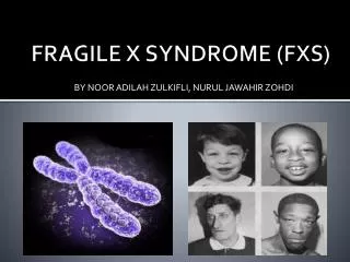
Fragile X syndrome. X linked disease, 1:4000 in males Clinical manifestations Cognitive difficulties Attention and behavioral problems Macro-orchidism Mild facial dysmorphologies Connective tissue abnormalities Anticipation. Fragile X chromosomes. Fragile X chromosomes.

E N D
Fragile X syndrome • X linked disease, 1:4000 in males • Clinical manifestations • Cognitive difficulties • Attention and behavioral problems • Macro-orchidism • Mild facial dysmorphologies • Connective tissue abnormalities • Anticipation
FRAXA- rare folate-sensitive fragile sites: mutation stages • At the loci of fragile sites there are naturally occurring polymorphisms of the number of copies of tandem repeats of the trinucleotide repeat CGG. • The fragile site is seen cytogenetically • The gene associated with the repeat is apparently normally expressed. • Beyond the premutation is the full mutation where the fragile site is seen and the relevant gene is transcriptionally silenced. • Normal chromosomes 5-55 copies of CGG • Premutation ~55-230 copies of CGG • Full mutation >~230 copies of CGG
The mutation - (CGG)n • In exon 1 - 5’ untranslated region. • Transmitting males • Normal alleles - 6-53, full mutation >230 repeats, • However there is a gray zone • Punctuations by AGGs at the 3’ end: (CGG)8-15 AGG (CGG)9-13 AGG(CGG)x • In very unstable alleles the 2 AGG are lost • 33 or less uninterrupted CGGs - stability, • >39 uninterrupted CGGs - instability
The FMR protein (FMRP) • The FMRP protein probably forms ribonucleoprotein (RNP) complexes, has 3 RNA-binding motifs, a RGG-box and 2 KH-domains. • Has a key role in regulation of translation. • Binds a subset of brain mRNAs including its own. • In brain, it shuttles a subset of mRNA to the dendritic spines. • Absence of the protein (deletion) cause immature dendritic spine morphology.
Fragile X syndrome - consequences of expansion • Methylation of the C (CGG) - due to mispaired Cs in secondary structures, are templates for methylation. • Methylation of the promoter is accompanied (reason unknown), leading to lack of transcription initiation. Disease mechanism -protein loss of function
Repeat expansion diseases • trinucleotide diseases: CGG, CAG and CTG • tetranucleotide - MD type 2, CCTG in intron 1. • pentanucletodie - SCA10, ATTCT up to 4,500 repeats in intron 1.
Repeat location Coding disorders Diseases with a CAG expansion within the coding region, produces an enlarged polyglutamine tract Huntigngton, Kennedy, Spinocerebellar ataxia type 1…) . Non coding disorders 1.Untranslated 5’ (Fragile X,syndrome, Spinocerebellar Ataxia type 12..) 2. Untranslated 3’ (myotonic dystrophy) 3. Intron (Friedreich ataxia, )
Features of Diseases caused by repeat instability • 40 neurodegenerative and neuromuscular diseases are caused by gene-specific instability of certain repeat tracts. • Most diseases are caused by triple repeat expansion. Some by longer repeated unit. • The common triplets are: (CTG)* (CAG) found in at least 14 diseases, (CCG)*(CGG), GCG*CGC, GAA. • Not all possible triplets are found to be expanded in these diseases. Only those that have the ability to form unusual DNA structures.
CAG expansion in the coding sequence • Huntington’s disease • Spinocerebellar ataxias • Kennedy’s disease
Gain of function at the protein level • The most common trinucleotide repeat causing disease by altering protein physiology is the (CAG)n. • (CAG)n in the coding region of a gene. Although expansion sizes, structures, cellular localization and functions of the resulting proteins differ, all (CAG)n-induced diseases are neurodegenerative disorders. • All disorders are associated with neuronal aggregates that contain the disease-causing gene product. • PolyQ stretches have an inherent ability to aggregate. Apart from binding to many other proteins, glutamine also shows self-interaction. • Once polyQ stretches exceed a certain length, they are no longer soluble and form aggregates.
Gain of function at the protein level • The threshold length, above which in vitro aggregation takes place, is strikingly similar to the threshold that causes disease, (40 CAGs). • Although it is tempting to hold the aggregates responsible for the development of disease, some evidence exists that the large aggregates are not the primary cause of cell toxicity.
Features of Diseases caused by repeat instability • Dynamic • Repeat tract length – correlates with age of onset and disease severity. • Anticipation - The number of inherited repeats increases significantly from generation to generation causing earlier onset and faster progression • Longer repeats – more likely to undergo expansion. • In many diseases (CAG repeats, glutamine) there is a late onset.
Hypothesis • The extended glutamine portion has a gain of toxic function which leads to a cumulative damage in the affected cells, possibly in the form of glutamine aggregate formation. • A new mechanism was recently suggested.
A universal mechanism • Onset and progression of the disease are determined by the rate of expansion of the tri-nucleotide repeat in certain cells in the patient’s body. • The disease manifests when the trinucleotide repeat expands beyond a certain threshold in a sufficient number of these cells and progresses as more and more cells do so. Kaplan et al, 2007
Friedreich’s Ataxia • The most common heritable form of ataxia associated with progressive gait and limb ataxia • Degeneration of large sensory neurons • Death is due to cardiac failure • Autosomal recessive,thus no anticipation • FRDA gene encoded frataxin • In FRDA disease the protein levels are severely reduced causing mitochondrial dysfunction
Friedreich’s Ataxia • The disease is caused by GAA-CTT repeat expansion • The repeat is located in intron 1, within an Alu sequences of the FRDA gene • Normal alleles 7-34 repeats • Disease causing >100 repeats • GAG interruptions • Very low mRNA levels
Model • Transcription-coupled triplex formation • The triplex blocks progress by RNA polymerase • No transcripts are produced • No protein • Loss of function
Disease Mechanisms • Loss of function at the protein level (fragile X) • Gain-of-function at the protein level (Huntington) • Loss of function at the RNA level (Friedreich’s Ataxia( • RNAgain of function (DM)
Myotonic dystrophy type 1 • AutosomalDominant inheritance • The most common form is adult muscular dystrophy, 1:8000 births • A wide spectrum of clinical phenotype: effecting the skeletal muscles (myotonia), eye, heart, endocrine systems,and more • CTG in the 3’ untranslated region of the DMPK gene in 98% of the cases • The remaining 2% are DM type 2 and found to carry a CCTG expansion in the ZNF9 gene.
Myotonic dystrophy type 1 • 5-27 repeats in normal alleles • Several thousands (>3,000) in patients • Variable phenotype and the anticipation are corrleated with the number of expanded repeats • The DMPK protein is a serin threonin protein kinase expressed exclusively in muscle and heart • Autophosphorylation and phosphorylation of Histon H1 and many other proteins such as myosin phosphatase. • DMPK phoshorylation inhibits the mypt1 activity -> high levels of the phos protein -> Ca+ sensitization of smooth muscles and cytoskeleton changes in non-muscle cells.
Myotonic dystrophy type 1 • The mutation cause dysfunction of of DMPK activity. • Suggestions: haploinsufficiency • However, mouse models did not support it: • 1. Heterozygous and homozygous DMPK knockout mice showed no myotonia (the major symptom) nor catarct characteristics of DM1. • 2. However, these mice developed late-onset skeletal myopathy and altered calcium ion homeostasis. • 3. Cardiac phenotype • Conclusion: haploinsufficiency (loss of function) plays a role but inactivation of the DMPK alone cannot be responsible for the complete DM1 phenotype.
Myotonic dystrophy type 1 • The expanded repeat containing mRNAs were retained in nuclear foci. • This is thought to occur because expanded RNA (CUG)n molecules. • Form secondary structures such as hairpins. • In the foci, RNA molecules sequester RNA-binding proteins (RNA-BPs), which bind specifically to (CUG)n (CUG-binding proteins (CUG-BPs). • Since mRNA processing, including splicing, is normally regulated by a dynamic complex of RNABPs, the function of these proteins in the presence of expanded (CUG)n was investigated.
Myotonic dystrophy type 1 • MBNL1 is a specific CUG-BP. • Proteins that are essential in terminal differentiation of muscle and photoreceptor cells. • It accumulates in the nuclear foci in DM1 cells, so that MBNL1 cannot exert its normal function during a critical period of cell differentiation.