Download
1 / 28
310 likes | 609 Views
Cardiovascular disease and the eye. Leo Semes, OD Professor, Optometry UAB, Birmingham, AL. Case Report. 75 WM Reports the following “At church last week it looked like I was seeing through cracked glass.” What additional information do you want from this history?.

E N D
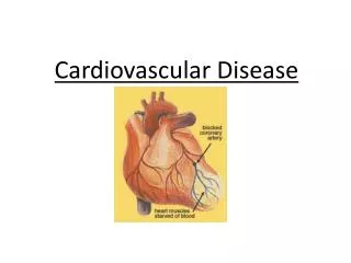
Cardiovascular disease and the eye Leo Semes, OD Professor, Optometry UAB, Birmingham, AL
Case Report • 75 WM • Reports the following “At church last week it looked like I was seeing through cracked glass.” • What additional information do you want from this history?
Cracked glass vision in a 75 WM One episode About 20 min Left eye only None; what might you expect? • Has it happened before / since? • How long did it last? • Was it one eye or both? Which one? • Did you experience any other symptoms?
Cracked glass vision in a 75 WM • What testing would you do? • VA • Anterior segment evaluation • Tonometry • Fundus evaluation • Results: All unremarkable for age
Cracked glass vision in a 75 WM • What could be the cause? • Local (vitreous traction) • Vascular • Retinal vasculature • Ophthalmic artery • Carotid artery • Vertebral basilar • Other?
How would you manage this patient? • Further testing? • Carotid auscultation • Ophthalmodynamometry • Additional carotid evaluation • General physical / vascular assessment • Results:
Blood supply to the eye academic.sun.ac.za/neurology/lectures/eyes98/sld030.htm
Blood supply to the eye Branches of CA: a = internal carotid artery b = vertebral artery c = basilar artery d = ophthalmic artery e = anterior cerebral artery f = middle cerebral artery g = posterior cerebral artery.
Cracked glass vision in a 75 WM • Outcomes • Carotid Doppler performed • Demonstrated > 90% blockage on left side • Patient recommended for L carotid endarterectomy • Successfully performed X 2 days • Patient survived an additional 7 years (succumbing to emphysema)
Floaters and sparkles • 63 W/M • “When I was grilling on July 4, I noticed sparks and floaters in my left eye.” • “I thought it was time for a CL check, so I came in to see you” • Sudden onset • No other symptoms
63 W/M with sparklers VA 20/20 in each eye Anterior segment evaluation – unremarkable for age DFE . . . (OS)
Outcomes • Sent to Internist for evaluation • Complained of dizziness to Internist • Carotid Doppler performed • Sufficient blockage to recommend carotid endarterectomy • Done within 3 weeks of visit to UABSO • Successful procedure
Other adverse outcomes of interrupted blood supply to the eye Central / branch retinal artery occlusion Ischemic optic neuropathy
Local arterial obstruction Partial, with hemi-field defect; total = sudden painless loss of vision (permanent)
Review - Retinal Emboli Emboli are blood clots or clumps of cholesterol and fatty material that break off from atherosclerotic plaques. When emboli lodge in blood vessels in or close to the eye, the eye's blood supply can be suddenly blocked. Emboli most often come from arteries in the chest or neck, but they can also come from the heart.
Retinal Emboli Emboli are a common cause of sudden but temporary vision loss; they can also cause permanent vision loss. Vision loss from emboli is sometimes described as a slow dimming of light or as a window shade being pulled down or up over the eye. When emboli travel to the brain and the eye at the same time, vision loss may be accompanied by loss of speech or weakness in an arm and leg. If these symptoms last more than a day, they indicate that the person has had a stroke. Diagnosis of the source of retinal emboli is done using ultrasonography or magnetic resonance angiography. Echocardiography and recordings of heart rhythm may be performed to determine if the person is at risk for further emboli.
Retinal Emboli Treatment may involve surgery (carotid endarterectomy) if test results show that the emboli may have come from the arteries in the neck and if the arteries are significantly narrowed. Otherwise, aspirin or other anticoagulants (sometimes called blood thinners) are used. Warfarin is given if test results show that emboli may have come from the heart. Treatment of atherosclerosis is important as well.
Review - Ischemic Optic Neuropathy Ischemic optic neuropathy is a sudden painless loss of vision in one eye from insufficient blood flow to the optic nerve. The cause is unknown. Atherosclerosis, diabetes, and high blood pressure may increase the risk of developing ischemic optic neuropathy. Temporal arteritis is a treatable form of ischemic optic neuropathy.
Review - Ischemic Optic Neuropathy Some people have pain or discomfort around the eye. An eye doctor diagnoses the condition by examining the eye. No proven treatments are available for most forms of ischemic optic neuropathy. For some people, vision improves without treatment. Only a small percentage of people experience the same symptoms in the other eye. Control of risk factors for atherosclerosis may help prevent ischemic optic neuropathy. This may be related to Sleep Apnea Syndrome (SAS)
Review - Ischemic Optic Neuropathy People with optic neuropathy due to temporal arteritis experience vision loss, which may be sudden in one eye. They may also experience headache, scalp tenderness at the temple, fever, and jaw pain when chewing. A doctor diagnoses the condition by examining the eye, performing blood tests, and performing a biopsy of the temporal artery. Treatment involves use of corticosteroids, mainly to prevent occurrence of disease in the other eye, but also to reduce risk of further vision loss in the affected eye.