Download
1 / 24
240 likes | 423 Views
Genetics. Cell Division Reference: Chapter 5. Principles of Cell Division. Cell organization Cell Nucleus Chromosomes Genes DNA. Types of Cell Division. Mitosis Occurs throughout life Functions growth, development, repair

E N D
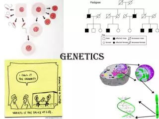
Genetics Cell Division Reference: Chapter 5
Principles of Cell Division • Cell organization • Cell Nucleus Chromosomes Genes DNA
Types of Cell Division • Mitosis • Occurs throughout life • Functions • growth, development, repair • Nuclear division in which chromosome number stays constant • Meiosis • Only occurs in reproductive organs • Reduces chromosome number by half
Vocabulary Review • Chromatin • Genetic material is long, thin strands dispersed throughout the nucleus in a tangled, fibrous mass. • Individual chromosomes all mixed together • Chromatin condenses and separates into individual chromosomes before mitosis begins. • Chromosomes • condensed rod-shaped DNA molecules • Only form during cell division
Vocabulary Review cont’d • Diploid (2N) number • characteristic chromosome number, chromosomes in pairs • Found in somatic cells • Haploid (N) number • half the diploid number, • found in gametes
Mitosis – Overview • DNA replication produces duplicated chromosomes • Composed of • 2 sister chromatids • Genetically identical • held together by a centromere • Centromere divides • Each chromatid becomes a daughter chromosome
Mitosis - Overview • Fig 5.3
Mitosis: Stages • Prophase • Nuclear membrane disappears, centrosomes migrate, spindle fibers appear • Metaphase • chromosomes line up at equator, associated with spindle fibers • Anaphase • centromeres divide, sister chromatids migrate to opposite poles, cytokinesis begins • Telophase • nuclear membranes form, spindle disappears, cytokinesis occurs
Meiosis- Overview • 2 divisions, 4 daughter cells • Cells are diploid at beginning of meiosis • Human body cells have 46 individual chromosomes • Chromosomes can be arranged into a set of 23 matched pairs called homologues • Similar in shape, size, and have genes which deal with the same traits.
Meiosis- Overview cont’d • Meiosis I • Homologues line up side by side at equator-synapsis • When pairs separate, each daughter cell receives one member of the pair • Cells are now haploid
Meiosis- Overview cont’d • Fig 5.9
Meiosis- Overview cont’d • Meiosis II • No replication of DNA occurs in this division • Centromeres divide and sister chromatids migrate to opposite poles to become individual chromosomes • Each of the four daughter cells produced has the haploid chromosome number and each chromosome is composed of one chromatid
Meiosis I - Stages • Prophase I • Synapsis occurs, nuclear membrane breaks down • Homologues line up side by side and crossing over occurs • Crossing over-exchange of segments of DNA between homologues • Crossing over-exchange of segments of DNA between homologues • Independent assortment of chromosome pairs
Independent alignment • Fig 5.11
Synapsis and crossing over • Fig 5.10
Meiosis I – Stages cont’d • Metaphase I • Homologous pairs line up at equator such that maternal or paternal member may be oriented toward either pole • Anaphase I • Homologous chromosomes (each still consisting of 2 chromatids) undergo independent assortment into daughter cells • Telophase I • Cytokinesis produces 2 daughter cells which are haploid • Interkinesis-period between meiosis I and meiosis II
Meiosis I in animal cells • Fig 5.12
Meiosis II - Stages • Prophase II • Cells have 1 member of each homologous pair • Metaphase II • Chromosomes line up at the equator • Anaphase II • Centromeres divide and daughter chromosomes migrate • Telophase II • Nuclei form, cytokinesis
Meiosis II • Fig 5.13
Meiosis in Humans • Spermatogenesis • Occurs in seminiferous tubules of testes • Begins at puberty and continues throughout life • Each meiotic division produces 4 haploid cells • Generates small cells, no organelles, only 1N nucleus
Meiosis in Humans cont’d • Oogenesis • Begins in vitro, primary oocytes present at birth • At puberty hormones stimulate 1 primary oocyte to complete the first meiotic division each month. • Each meiotic division produces only 1 haploid egg
Meiosis in Humans • Oogenesis: cont’d • Cell division is asymmetrical • Majority of cell contents stay in one half, will eventually develop into ovum, containing all necessary organelles for all future cells. • Smaller cell, polar body, degenerates and disappears. • Second meiotic division not completed unless egg is fertilized by sperm.
Spermatogenesis and oogenesis • Fig 5.16