Download
1 / 61
680 likes | 1.2k Views
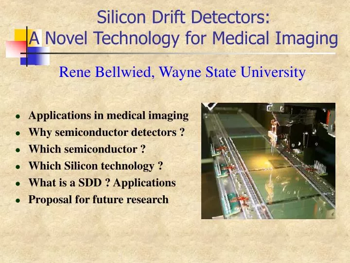
Silicon Drift Detectors: A Novel Technology for Medical Imaging. Rene Bellwied, Wayne State University. Applications in medical imaging Why semiconductor detectors ? Which semiconductor ? Which Silicon technology ? What is a SDD ? Applications Proposal for future research.

E N D
Silicon Drift Detectors: A Novel Technology for Medical Imaging Rene Bellwied, Wayne State University • Applications in medical imaging • Why semiconductor detectors ? • Which semiconductor ? • Which Silicon technology ? • What is a SDD ? Applications • Proposal for future research
Why Semiconductors ? • Presently most medical devices are based on photo-imaging (film) => excellent resolution but low sensitivity and lack of image uniformity => long exposure times in b- and x-ray imaging and development time. • New idea: single particle counting using semiconductor detectors has the following advantages: • High sensitivity (low exposure time) • High dynamic range and excellent linearity • Energy discrimination of particles • Direct digital imaging and online image display • Very good resolution (< 50 mm) • Lately, digital imaging based on integrating devices (i.e. MWPC (multi-wire proportional chambers, gas devices). Sensitivity better than film but resolution poorer (~400 mm)
Medical Applications X-ray applications • Digital mammography <E> = 20 keV • Dental X-ray tube <E>= 35 keV • Fast frame medical diagnostics Nuclear medicine • Thyroid measurements, <E> = 60-140 keV photons • DNA probe array, b-emitter (32P, 14C, 35S), <E> = 50-700 keV
X-ray Mammography statistics • Breast cancer • Most frequent neoplastic pathologies in women in the world • One of the first causes of death. U.S. has one of the highest rates in the world. • Last year 180,000 women in the U.S. were diagnosed, 44,000 women died of breast cancer • During last 50 years incidence rate (probability of woman infected during life cycle) is increasing • In 1950 the probability was one in fifty (0.2%) • Last year the rate was one in eight (12.5%) • Mammography • 85-90% malignancies are visible on mammography • Early diagnosis • 5-year survival rate after treatment is over 95% for localised breast cancer (dimensions less than 15 mm) • 5-year survival rate after treatment is less than 20% for metastatic cancer • Life quality improved: conservative surgery • Mammographic screening • Effective tool in reducing breast cancer mortality for women aged 50 years and over • Benefits for women younger than 50? Presently not applied because of dose.
Dose Considerations • Defining factors • Mammography relies on photon attenuation coefficient differences between tumours and normal breast tissue • The highest contrast between micro-calcification and glandular tissue is at low photon energies (generally below 30 keV) • But low energies have small photon transmission coefficients as a function of tissue thickness. • Breast compression leads to large improvements in dose rates. • Young vs. old women • single shot doses are low (less than 1 mGy), but dose integration over several years increases risk of associated carcinogenesis. • Young women have denser breasts, which strongly reduces the contrast to cancerous spots. Higher exposures with higher doses have to be used. • Breast cancer is most dangerous in young women as it tends to develop faster that in older patients. • The main proposed way to reduce the dose is to significantly increase the image quality
Definitions for device comparisons • Optical Density (O.D.) • The optical density of a film system can be directly compared to the count response for a digital system. • The optical density is a measure of the ‘blackening’ of a mammographic film • Spatial frequency • Spatial frequency n is measured in lp/mm (line pairs per mm) • Definition: n = 1 / (2 spatial resolution), e.g. 1 / 0.2 = 5 lp/mm for 100 micron resolution. In a pixel system the pixel size should be at least twice as small as the highest detectable spatial frequency. • Small objects require sharp images i.e. high spatial frequencies • Highly granular systems have a more stable MTF with increasing spatial frequency. • MTF is being measured at a fixed spatial frequency for comparison
MTF and DQE measurements • Modulation Transfer Function (MTF): • Spatial resolution properties of the imaging system • LSF(x) Line Spread Function: image of an “infinitely thin” line object • Detective Quantum Efficiency (DQE): • Noise transfer properties through the different stages of the imaging system
Digital Mammography (DM) • Main features • Linear response with X-ray exposure • Wide dynamic range (104 – 105) • Mammography of dense breast • Reduced radiation dose • Exposure determined as a function of the Signal to Noise Ratio (SNR) not of the Optical Density of the film (OD) • Dose reduction from 20 to 80 % • Image processing • time required for the examination (texp<1s; Tproc~minutes) • Limitations (?) • Spatial resolution • Film-screen systems 20 lp/mm • Digital systems 5 lp/mm (spatial frequency is smaller, but image is sharper) • Monitor resolution • Monitor 2000x2500 pixels, resolution 0.1 mm
Development of digital imaging • Need to be superior to standard X-ray film cassette: • Better resolution • Lower radiation dose, shorter exposure time • Need to match film detector size (18 x 24 cm2) • Problem: quantum efficiency of 300 mm Si wafer for medical x-rays is at best 25 %. Use Silicon for readout but not for signal generation. • Development based on amorphous Si (a-Si): detector based on converter with a-Si active matrix. • Indirect systems: scintillator as converter (e.g. CsI or P) , active matrix records visible photons from scintillator • Direct systems: use heavy semi-conductor as converter (e.g. a-Se, GaAs) with direct charge transfer to active matrix
Direct conversion of photons Minimises image blurring and avoids an extra conversion stage from X-rays into visible light Higher SNR with respect to phosphor-based systems Higher spatial resolution Single Photon counting More efficient noise discrimination Higher SNR for polychromatic beams Direct Digital Mammography (DMM)
Amorphous vs. crystalline semiconductors • Advantage of amorphous semiconductors: Can be produced into any size detectors (i.e. a-Si can match film detector size) • Disadvantage of amorphous semiconductors: Charge carrier lifetimes are orders of magnitude lower than in crystalline semiconductors. High voltages have to be applied to collect charges fast.
Famous recent commercial products • GE Senograph 2000D flat panel device: • Indirect system based on a Phosphor scintillator converter and a-Si matrix for readout (first digital device with FDA approval (2000)). • Fischer and Siemens slot scanning devices • Indirect systems based on Phosphor screen and CCD array. • AGFA-Gevaert flat panel device: • Direct system based on a-Se converter with a-Si matrix for readout.
Commercial systems for DM • Photostimulable phosphors (Imaging Plates) • Fujifilm HR-V 18 x 24 cm2 • pixel 100 x 100 mm2 • Double read-out • Flat Panels • GE Senographe 2000D • Revolution ™ Flat Panel Digital Detector • 18x 24 cm2, pixel 100 x 100 mm2 • 11 years R&D and 130 M$ investment • I Digital Mammographic system approved by FDA (2000) • Slot scanning systems • Fischer Imaging SenoScan • Phosphor screen +OF taper+ CCD array • 22 x 30 cm2 in 4 s • Pixel 50 x 50 mm2 • 10 years R&D and 30 M$ investment • Approved by FDA (2001)
Photon Counting Chip (PCC) VLSI Circuit developed at CERN in the frame of the MEDIPIX European collaboration CERN, Universities and INFN Pisa and Napoli (IT), Universities of Glasgow (UK) and Freiburg (GE) http://medipix.web.cern.ch/MEDIPIX/ EP CERN SACMOS 1 mm FASELEC now Philips AG, Zürich Pixel 170 x 170 mm2 channels 64 x 64 Area 1.7 cm2 Threshold adjust 3-bits Pseudo-random counter 15-bits Read-Out and Config. Freq. 10 MHz Read-Out Speed 400 ms/image 12 mm
Photon counting vs. optical density Optical density as a function of dose for mammographic film Photon counting as a function of dose for digital device 4.0 10000 1.0 2000 10 20 30 40 50 60 70 80 mAs 0.0 0.5 1.0 1.5 2.0 2.5 3.0 log(E/E0) =10 mAs =40 mAs
GaAs PCC Mammographic system Radiographic system Flat panel (a-Si+CsI) Fluoroscopic system MTF Comparison (according to MEDIPIX study) Direct detection of the photons Improved spatial resolution
GaAs PCC Trixell Fluoroscopic system Mammographic system Radiographic system DQE Comparison (according to MEDIPIX study) DQE of the pixel detector is higher and more stable with the frequency Due to direct detection of photons and Single photon counting Improved noise transfer properties
Which Semiconductors ? • Generally available: Ge, Si, GaAs, CdTe • Ge needs liquid nitrogen cooling to yield good resolution • GaAs and CdTe have higher X-ray absorption efficiency than Si in relevant range (<E>=10-70 keV) At 20 keV the detection efficiency for a 200 mm thickness GaAs layer is 98%, which is four times higher than the equivalent efficiency in Silicon. • GaAs is more advanced than CdTe but both technologies are in their prototype stages compared to Silicon.
Performance Comparison Si (left) vs. GaAs (right) • C.Schwarz et al., NIM A 466, 87 (2001)
Relevant Literature • S. Webb, “The Physics Of Medical Imaging”, Institute of Physics Publishing (1998) • M. Sandborg and G.A. Carlsson, “Influence of x-ray energy spectrum, contrasting detail and detector on the SNR and DQE in projection radiography”, Phys.Med.Biol. 37 (1992) 1245 • H.J.Besch, “Radiation Detectors im medical and biological applications”, NIM A419 (1998) 202 • S.R.Amendolia et al, “Spectroscopic and imaging capabilities of a pixellated photon counting system”, NIM A466 (2001) 74 • C.Schwarz et al., “Measurements with Si and GaAs pixel detectors bonded to photon counting readout chips”, NIM A466 (2001) 87 • A.Castoldi et al., “The Controlled Drift Detector: a new detector for fast frame readout X-ray imaging”, NIM A461 (2001) 405 • A Castoldi et al., “The Controlled Drift Detector”, NIM A439 (2000) 519
Phantom 110 cm Detector Imaging of mammographic phantoms Lucite cylinder thickness 4 cm diameter 10 cm Al disks diameter 4 mm Embedded in wax cylindersdiameter 12 mm
Image comparison Si 300 mm 0 GaAs 200 mm Film 12 bits 170 mm 255 125 mm 100mm 75 mm 40 mm 25 mm Exposure = 32 mAs Exposure Time ~ 1 s Dose = 6 mGy 10.9 mm
. . . . . . . . . . . . . . . . . . RMI 156 phantom • American College of Radiology (ACR) Accreditation phantom • RMI 156 features • Details • 6 Nylon fibers • Ø 1.56 – 0.4 mm • 5 microcalcification groups • Ø 0.54 – 0.16 mm • 5 tumour masses • 2.00 – 0.25 mm • Dimensions 4.5 x 10.2 x 10.8 cm3 • Compressed breast 4.2 cm thick
Pixel detector 6x6 scanning Pixel 170 mm Entrance Dose/image = 4 mGy Mammographic film Digitized 100 mm pitch 12 bits Entrance Dose = 4 mGy Images 255 6 cm 0 6 cm
Dose comparison • Al disk, 125 mm thick, embedded in 3 mm thick wax cylinder, Lucite block 4 cm thick • Images • Dimension 1.2 cm2 • Pixel 170 mm 0 255 Film 32 mAs GaAs 8 mAs Dose ~ 6 mGy Dose ~ 1.5 mGy
Why Silicon Drift Detectors ? • Generally available: CCD, Si pixels, Si strip, Si drift • CCD (charge-coupled devices) are slow and not very radiation resistant. • Silicon pixels are fast and have high resolution but they are very expensive and the connection to the electronics (bump bonded) is a difficult technical process, but the main electronics development (PCC) is optimized for bump-bonding • Silicon strip detectors have relatively poor resolution and are not cost competitive to drift detectors.
Silicon Drift Detector Characteristics • Large units can be mass produced (presently up to 10 by 10 cm) • Detector operates well at room temperature or with Peltier cell cooling • Electronics density is order of magnitude smaller than resolution. (electronics pitch = 250 mm, position resolution < 20 mm) • Three dimensional point determination with one dimensional readout.
Present status of technology STAR (detector at Relativistic Heavy Ion Collider (RHIC) at BNL on Long Island) • 4in. NTD material, 3 kWcm, 280 mm thick, 6.3 by 6.3 cm area • 250 mm readout pitch, 61,440 pixels per detector • SINTEF produced 250 good wafers (70% yield) ALICE (future detector at Large Hadron Collider (LHC) at CERN in Geneva, Switzerland) • 5in. NTD material, 2 kWcm, 280 mm thick, 280 mm pitch • CANBERRA produced around 100 prototypes, good yield Future (potentially for detector at Next Linear Collider (NLC) in ?) • 6in. NTD, 150 micron thick, any pitch between 200-400 mm • 10 by 10 cm wafer • low radiation length, low cost for large area
STAR/SVT at RHIC (BNL) • Search for the quark-gluon plasma (QGP) and investigate the behavior of strongly interacting matter at high energy density. Installed in February 2001, first beam in July 2001. 2500 charged particles/event Radiation length: 1.4% per layer • 0.3% silicon, 0.5% FEE (FrontEnd Electronics), • 0.6% cooling and support. Beryllium support structure. • FEE placed beside wafers. Water cooling. • SVT costs $7M for 0.7m2 of silicon.
RHIC Au-Au Beam Collisions Approach Collision Particle Shower
The SVT in STAR Construction in progress Connecting components
STAR-SVT characteristics • 216 wafers (bi-directional drift) = 432 hybrids • 3 barrels, 103,680 channels, 13,271,040 pixels • 6 by 6 cm active area = max. 3 cm drift • 3 mm (inactive) guard area • max. HV = 1500 V • max. drift time = 5 ms, (TPC drift time = 50 ms) • anode pitch = 250 mm, cathode pitch = 150 mm
The SVT in STAR The final device…. … and all its connections
R&D on SDD thickness All recent silicon detectors were produced from 300 mm thickness wafers
Mature technology. <10 micron resolution achievable with $’s and R&D. Easy along one axis (anodes). <0.5% radiation length/layer achievable if FEE moved to edges. Low number of channels translates to low cost silicon detectors with good resolution. Detector could be operated with air cooling at room temperature Technology is viable for position and energy measurements and first tests show excellent response to single photons. STAR-SDD Summary
We have an agreement with MEDIPIX to use their photon counting chip in connection with our SDD devices. An R&D program to convert bump-bonded chips to wire-bonded chips has started. Where do we go from here ? • Strong European efforts are already underway, based on state of the art high energy semi-conductor detectors • MEDIPIX collaboration at CERN is an EU supported project • IMI collaboration is an Italian consortium of research groups and industry
Integrated Mammographic Imaging Project • The aim of the project is to build an innovative system for both morphologic and functional mammography The project has been financially supported by the Italian Government The research fields are: • Monochromatic X ray tube for mammography • Digital mammographic system based on GaAs pixel detectors • Bump-Bonding technology for GaAs pixel detectors • High resolution mammoscintigraphic system • The project is managed by five Italian companies in collaboration with the Universities of Ferrara, Napoli, Pisa, Roma “La Sapienza” and the “Istituto Nazionale di Fisica Nucleare” (INFN) • 3 years and 4 MEuro investment
Form an Interdisciplinary Medical Imaging Project at Wayne State University (WSU-IMIP) between the Physics Department, the Medical School, and the Karmanos Institute Our short-term goals • Collaborate with MEDIPIX on the electronics and with Brookhaven National Laboratory on the Silicon Drift Detectors • Find a U.S. based industrial partner (Photon Imaging, Inc. ?) • Obtain funding through the WSU Life Science Corridor Initiative