Download
1 / 50
510 likes | 954 Views
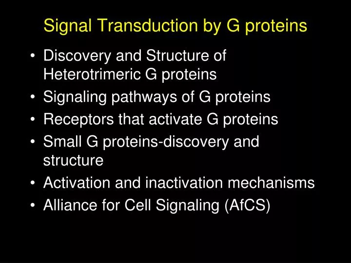
Signal Transduction by G proteins. Discovery and Structure of Heterotrimeric G proteins Signaling pathways of G proteins Receptors that activate G proteins Small G proteins-discovery and structure Activation and inactivation mechanisms Alliance for Cell Signaling (AfCS).

E N D
Signal Transduction by G proteins • Discovery and Structure of Heterotrimeric G proteins • Signaling pathways of G proteins • Receptors that activate G proteins • Small G proteins-discovery and structure • Activation and inactivation mechanisms • Alliance for Cell Signaling (AfCS)
Discovery of G proteins Martin Rodbell first proposed the concept of “discriminator-transducer-amplifier” to address the problem: “How can many hormones (epinephrine, ACTH, TSH, LH, secretin, and glucagon) activate lipolysis and cAMP production in adipocytes through presumably a single cyclase? He called this problem “too many angels on a pinhead.” His work identified GTP as important for the “transducer”. Nobel prize, 1994 His work was not initially received well by the scientific community:
Discovery of G proteins Al Gilman purified the first G proteins. His lab took advantage of S49 lymphoma cells that lacked Gsa (although at the time, the cells were thought to lack adenylate cyclase, thus the name cyc-). Reconstitution experiment rationale: Isolate membranes from cyc- cells, then add back fractions from donor wt membranes that restore adenylate cyclase activity. Nobel prize, 1994 Donor membranes were incubated for increasing time at 30oC, which inactivates the adenylate cyclase activity (- - - - -). Fortunately, G proteins are relatively heat stable. Addition of NaF, Gpp(NH)p, GTP, or GTP and isoproterenol restored activity in the cyc- membranes. Ross, et al. JBC (1978)
Adenylyl Cyclases as Coincidence Detectors AC Type: I II III V Gas GTP 0 ? 0 Ca2+/Calmodulin 0 0 Gbg 0 0 Protein kinase C 0 0 0 0 Gai GTP Gs and Gi have opposing actions on adenylyl cyclases Toxins help identify a second G protein. Both toxins result in increased cAMP production, but by different mechanisms. Cholera toxin ADP-ribosylates GaS, while pertussis toxin clearly did not act on the newly purified GaS (could use radiolabeled ADP). Using pertussis toxin to ADP-ribosylate the target, Gilman lab identified and purified Gai.
Signal Transduction by G proteins • Discovery and Structure of Heterotrimeric G proteins • Signaling pathways of G proteins • Receptors that activate G proteins • Small G proteins-discovery and structure • Activation and inactivation mechanisms • Alliance for Cell Signaling (AfCS)
G protein signal transduction Neves, Ram, Iyengar, Science 2002
Structure of G proteins Iiri, et al. NEJM (1999)
Hydrolysis of GTP • Arg & Gln stabilize the b and g phospates of GTP molecule in correct orientation for hydrolysis by H2O • Hydrolysis leads to major conformation change in Gsa • Mutations in the Gln or Arg (or ADP ribosylation by cholera toxin) blocks the ability to stabilize transition state, and therefore locks G protein in the “on” position. • Examples include adenomas of pituitary and thyroid glands (GH secreting tumors, acromegaly), and McCune-Albright syndrome. Iiri, et al. NEJM (1999)
Canonical Gs Signaling Pathway For interactive pathways at STKE: Gs pathway http://stke.sciencemag.org/cgi/cm/CMP_6634 Gi pathway http://stke.sciencemag.org/cgi/cm/CMP_7430 Gq pathway http://stke.sciencemag.org/cgi/cm/CMP_6680 G12 pathway http://stke.sciencemag.org/cgi/cm/CMP_8022 Neves, Ram, Iyengar, Science 2002
McCune-Albright Syndrome • Polyostotic fibrous dysplasia • Café au lait skin lesions • Autonomous hyperfunction of one or more endocrine glands • Gonadotropin-independent precocious puberty • Cushing’s syndrome • Acromegaly The constellation of symptoms varies from one individual to the next. How can a single mutation present in patches?
Testotoxicosis and PHP, 1a • Two unrelated boys with both gain-of function and loss-of function diseases associated with Gs. • Testotoxicosis=inappropriate secretion of testosterone. Usually under the control of LH (luteinizing hormone) secretion by the pituitary. LH receptors in the testes activate Gs. • Pseudohypoparathyroidism=lack of PTH (parathyroid hormone) signaling resulting in impaired calcium homeostasis and bone abnormalities (Albright’s osteodystrophy). PTH receptors in bone activate Gs. Mechanism?
Human Genome Sequencing More added complexity: Human Fly Worm Yeast Plant
Signal Transduction by G proteins • Discovery and Structure of Heterotrimeric G proteins • Signaling pathways of G proteins • Receptors that activate G proteins • Small G proteins-discovery and structure • Activation and inactivation mechanisms • Alliance for Cell Signaling (AfCS)
G protein signaling • Many ligands • Robust switches • Multiple effectors • Conserved 7 TM architecture • More than 50% of drugs target GPCRs Bockaert & Pin, EMBO J (1999)
G protein-coupled receptors • 5 main families • Conserved 7 TM architecture
Identifying Ligands for Orphan GPCRS Big Pharm approach: set up individual stable cell lines expressing each orphan GPCR. Fractionate peptides, tissue factors, etc. and apply to each cell line. Example: Orexin receptors Cottage industry approach: expression cloning strategy in Xenopus oocytes. Use sib selection to identify cDNAs that encode desired receptor. Example: Thrombin receptor
New concepts for GPCR signaling Using mainly two-hybrid screening approaches, many proteins have been found to interact with portions of the GPCRs. Non-PDZ scaffolds: AKAPs (A-Kinase Anchoring Proteins, JAK2 (Janus Activated Kinase), homer, b-arrestins PDZ scaffolds: InaD, PSD-95 (Post-Synaptic Density), NHERF (Na/H Exchanger Regulatory Factor). The arrestins have been found to bind to other signaling proteins and activate downstream effectors: Examples: src, Raf & ERK, ASK1 & JUNK3 Lefkowitz reviews
Arrestins act as scaffolds for ERK and JNK signaling pathways Lefkowitz reviews
Bonus material--Dynamic scaffolding Visual system in the fly NinaD is scaffold protein that binds PKC, PLC, and TRP channel Crystal structure of PDZ5 reveals a disulfide bond . . . Does it occur in vivo and is it important? Mishra et al Cell 2007
Bonus material--Dynamic scaffolding Visual system in the fly Titrate the disulfide bond with increasing concentration of DTT Redox Potential of the disulfide in InaD is very strong Most cytosolic proteins are -0.23 to -0.30 Mishra et al Cell 2007
Light-dependent inactivation impaired Bonus material--Dynamic scaffolding Visual system in the fly Make transgenic fly with C645S mutation Do electrophysiology (inaD2= KO, inaDwt= WT rescue) Single photon response OK, but . . .
Bonus material--Dynamic scaffolding Visual system in the fly NinaD is scaffold protein that binds PKC, PLC, and TRP channel Crystal structure of PDZ5 reveals a disulfide bond . . . Does it occur in vivo and is it important? WT InaDC645S
Signal Transduction by G proteins • Discovery and Structure of Heterotrimeric G proteins • Signaling pathways of G proteins • Receptors that activate G proteins • Small G proteins-discovery and structure • Activation and inactivation mechanisms • Alliance for Cell Signaling (AfCS)
Signaling GTPases are Allosteric Switches Ras = classical “monomeric” GTPase Swi2 -phosphate g Swi1 Ras-GTP vs. Ras-GDP Binding g-phosphate changes the conformations of two small surface elements, called “switch 1 and 2” Discovery of Small G proteins Ras genes first identified in ‘60’s as transforming genes of rat sarcoma viruses. Weinberg, Varmus, Bishop and others in the early ‘80’s showed that many cancer cells have mutated versions of ras. Activated form of ras found in 90% of pancreatic carcinomas, 50% of colon adenocarcinomas, and 20% of malignant melanomas.
Rho/Rac/Cdc42 In early ‘90’s, Alan Hall discovered that newly characterized ras homologs (rho, rac, cdc42) induced cytoskeletal changes. Reviewed by Hall, Science 1998
Ras superfamily of small G proteins Takai, et al. Physiological Reviews, 2001
GTPases: How to use reverse genetics to identify their roles in cell regulation Depends on understanding how the machines work Epistasis question: Where in a pathway does a specific protein convey its particular message? C D E A B Response M N Q Idea: 1. Inhibit activity of the protein of interest 2. Increase activity of the protein of interest How to do this? Drugs, genetic diseases, mouse KOs, and . . .
Reverse genetics: express one or two mutant versions of the protein of interest Depends on understanding how the machines work 1. Inhibit activity of the protein with a “dominant-negative” interfering mutant of that protein The mutant titrates (binds up) a limiting component to block the normal protein’s signal 2. Increase activity of the protein with a “dominant-positive” or “constitutively active” interfering mutant of the protein The mutant exerts the same effect as the normal protein would, if it were activated in the cell
GEF GDP GTP GAP Reverse genetics: small GTPases as examples Depends on understanding how the machines work “Dominant-negative” mutation GEF “Dominant-positive” mutation GDP Binds GEF but cannot replace GDP by GTP; so GEF not available for activating normal protein Cannot hydrolyze GTP, so remains always active Pi The mutant titrates (binds up) a limiting component to block the normal protein’s signal The mutant exerts the same effect as the normal protein would, if it were activated
Reverse genetics: advantages/pitfalls of using dominant-interfering mutants Pro: Con: Quick-and-dirty; no biochem Dominant-negatives Over-expression can titrate too many proteins (or the wrong proteins Many different families of signaling proteins amenable . . . once we understand how one of them works Dominant positives Not always precise mimics of the normal protein (e.g., may be in the wrong place)) Examples: RTKs? Other kinases? Adaptors? Can induce adaptation, turn-off mechanisms Hard to apply to complex networks Therefore . . . Still need biochemistry
Ras Hierachy of small G protein activation Use of constitutively active or dominant negative mutant small G proteins revealed that ras and cdc42 can activate rac. Rac, in addition to inducing lamellipodia, also activates Rho. Takai, et al. Physiological Reviews, 2001
Rho/Rac/Cdc42 signaling in actin assembly Takai, et al. Physiological Reviews, 2001
Identification of RasGAP V12 McCormick injected Xenopus oocytes with oncogenic ras (V12) versus wt ras (G12) and monitored germinal vesicle breakdown (GVB) (top panel) % GVB G12 Then loaded ras with a-32P GTP, injected into oocytes, did immppt at increasing times and determined if GTP or GDP was bound (bottom panel) [ras] (ng) Rate of GTP hydrolysis is 300-fold faster in oocytes than in vitro! V12 % Ras-GTP Purified the factor that promoted GTPase activity, cloned and named it GAP (or ras-GAP). Another ras-GAP later identified is NF1 (the gene mutated in neurofibromatosis, i.e., Elephant Man Syndrome). G12 Time (min)
Signal Transduction by G proteins • Discovery and Structure of Heterotrimeric G proteins • Signaling pathways of G proteins • Receptors that activate G proteins • Small G proteins-discovery and structure • Activation and inactivation mechanisms • Alliance for Cell Signaling (AfCS)
Small G proteins “turn off” mechanisms RhoGAPs outnumber the small G proteins Rho/Rac/Cdc42 by nearly 5-fold. Why so much redundancy? Luo group did RNAi against 17 of the 20 RhoGAPs in fly. Six caused lethality when expressed ubiquitously. Tissue specific expression of RNAi revealed unique phenotypes. P190RhoGAP implicated in axon withdrawal. Increasing amounts of RNAi caused more axon withdrawal (panels C-G). Why so many RhoGAPs? Billuart, et al. Cell (2001)
Small G protein “turn on” mechanisms First mammalian GEF, Dbl, isolated in 1985 as an oncogene in NIH 3T3 focus forming assay. It had an 180 amino acid domain with homology to yeast CDC24. This domain, named DH (Dbl homology) is necessary for GEF activity. In 1991, Dbl shown to catalyze nucleotide exchange on Cdc42. Schmidt & Hall, Genes & Dev. (2002) Dbl= Diffuse B-cell lymphoma
Rho/Rac/CDC42 activation of downstream effectors Rho Effectors: PI 3-Kinase, PLD, Rho Kinase, Rhophilin, and others. Rac-interacts via a CRIB domain in downstream effectors. CRIB (Cdc42/Rac interacting binding) Effectors: NADPH oxidase, PAK, PI 3-Kinase, MLK2,3, POSH, DGK Cdc42 Effectors: PI e-Kinase, PAK, WASP, S6-Kinase, MLK2,3, Borg
The GTPase switch Schmidt & Hall, Genes & Dev. (2002)
Mechanism of GDI-rab association Ypt1 is a small G protein (rab family). Rab-GDI binds the GDP-Ypt and removes it from the PM. Recent co-crystal structure reveals possible mechanism. Rak, et al. Does this interaction really happen in cells? Probably--mutations in domain II cleft abolish ability of RabGDI to remove Ypt1 from PM.
Signal Transduction by G proteins • Discovery and Structure of Heterotrimeric G proteins • Signaling pathways of G proteins • Receptors that activate G proteins • Small G proteins-discovery and structure • Activation and inactivation mechanisms • Alliance for Cell Signaling (AfCS)
Central Questions of the AfCS: I Question 1: How complex is signal processing in cells? The set of ligands for cellular receptors is the potential combinatorial code of inputs. How much of this input complexity can a cell uniquely decode as outputs? Experiment: Systematic single- and double- (multi?) ligand screens. Classify output responses; determine degree of crosstalk; identify “hotspots” for later quantitative analysis. New Technologies: Analytic methods to classify and compare multi-dimensional data for different ligand combinations
Central Questions of the AfCS: II Question 2: What is the structure of the whole signaling network? Is the connectivity sparse or dense? Experiment: Wholesale mapping of relevant protein-protein and small molecule-protein interactions. New Technologies: High-throughput assays for intermolecular interactions in vivo, especially in response to ligand stimulation.
Central Questions of the AfCS: III Question 3: How much does network topology constrain signal processing capability? How much function is specified by the nature of the connections, rather than by the specific biochemical constants of individual activities. Experiment: Perturbation methods; gain and loss of function, coupled with functional assays. New Technologies: Perturbations in vivo, singly and in combinations.
Central Questions of the AfCS: IV Question 4: What are the dynamics of the signaling network? Can we visualize how information propagates through the network and emerges as functional activities? Question 5: Can functional modules be abstracted mathematically? Can we make physical models and predict input-output relationships Question 6: Why is the network the way it is? Why have the observed solutions been chosen? What is being optimized?