Download
1 / 26
260 likes | 411 Views
KAROTYPING CHROMSOME ABNORMALITIES. * Usually done from the 15th or 16th week to the 20 th week (2 nd trimester). *Only at this point in a pregnancy is there sufficient amniotic fluid to allow some of it to be drawn off without significant danger to the fetus.

E N D
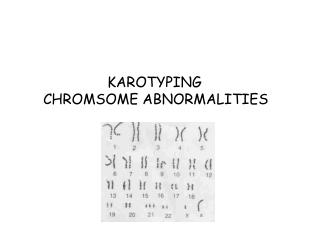
KAROTYPING CHROMSOME ABNORMALITIES
*Usually done from the 15th or 16th week to the 20th week (2nd trimester). *Only at this point in a pregnancy is there sufficient amniotic fluid to allow some of it to be drawn off without significant danger to the fetus. * Only about .5% risk of the test causing a miscarriage.
*In chorionic villi sampling (or biopsy), a small sample of chorioncells are collected. • Chorion is a membrane that develops around an embryo • and contributes to the formation of the placenta. • Done beginning in the 10th-12th week (1st trimester). • *Risk of miscarriage is at least 1-3%, or 2-6 times higher than with amniocentesis.
*A band is defined as that part of a chromosome which is clearly distinguishable from its adjacent segments by appearing darker or brighter with one or more banding techniques. *The chromosomes are visualized as consisting of a continuous series of bright and dark bands. *The banding techniques fall into two principal groups: 1) Bands distributed along the length of the whole chromosome (EX: G-banding) 2) Bands stained at a restricted number of specific sites. C-banding methods do not permit identification of every chromosome in the somatic cell, but can be used to identify specific chromosomes.
*G-bands are most commonly used. Shows bright and dark bands in an alternating fashion. *They take their name from the Giemsa dye. *Dark regions are heterochromatic. *Areas with a lot of adenine and thymine *Contains no functional genes *The bright regions are euchromatic. *Areas with a lot of guanine and cytosine *Contains functional genes (make proteins)
www.pathology.washington.edu/galleries/Cytogallery/cytogallery.htmlwww.pathology.washington.edu/galleries/Cytogallery/cytogallery.html
AUTOSOMAL ABNORMALITIES • The majority of human chromosomal abnormalities occur in the autosomes. • *Most common: • 1.Monosomy: one copy of a chromosome • --All fetuses spontaneously abort early in pregnancy. • 2. Trisomy: 3 copies of a chromosome • --Almost all fetuses die before birth
*1 in 800 live births *Approximately 85% spontaneously abort. *About 2-4% (of 350,000) are genetically mosaic. --Some of their cells have chromosome 21 trisomy while others do not, resulting in generally milder symptoms
Characteristic clenched hand Rocker bottom feet
SEX CHROMSOMAL ABNORMALITIES • *In frequency of occurrence, they are only slightly less common than autosomal abnormalities. • They are usually much less severe in their effects. • The high frequency of people with sex chromosome aberrations is partly due to the fact that they are rarely lethal conditions. • * Sex chromosome gross abnormalities can be diagnosed before birth by amniocentesis and chorionic villi sampling.
KLINEFELTER’S SYNDROME • 1 in 500 and 1 in 1000 male births • *Genotype is XXY or more rarely XXXY, XXXXY • *Relatively high-pitched voices (low levels of testosterone) • *Asexual to feminine body contours as well as breast enlargement (low levels of testosterone) • *Comparatively little facial and body hair. • *Sterile or nearly so
“SUPER-MALE” • *It may be as common as 1 in 900 male births to as rare as 1 in 1500 or even 1 in 2,000. • *Genotype is XYY • Usually tall (above 6 feet) and generally appear and act normal • During adolescence, they often are slender, have severe facial acne, and are poorly coordinated • *Some researchers suggest that the high testosterone levels can make them somewhat more prone to violence and that this may cause higher rates of wife beating.
TURNER’S SYNDROME • *Genotype is XO • 1 in 3,000 female infants to 1 in 5,000 • 4 foot 7 inches as adults, distinctive webbed necks (i.e., extra folds of skin), small jaws, and high arched palates. • *If diagnosed in early childhood, regular injections of human growth hormones can increase their stature by a few inches. • *Beginning around the normal age of puberty, estrogen replacement therapy can result in some breast development and menstruation.