Download
1 / 1
10 likes | 123 Views
Case of the week – 06-12: Coarctation, MS and bicuspid AoV. Clinical history: A 17-yr-old ♀, referred with hypertension.

E N D
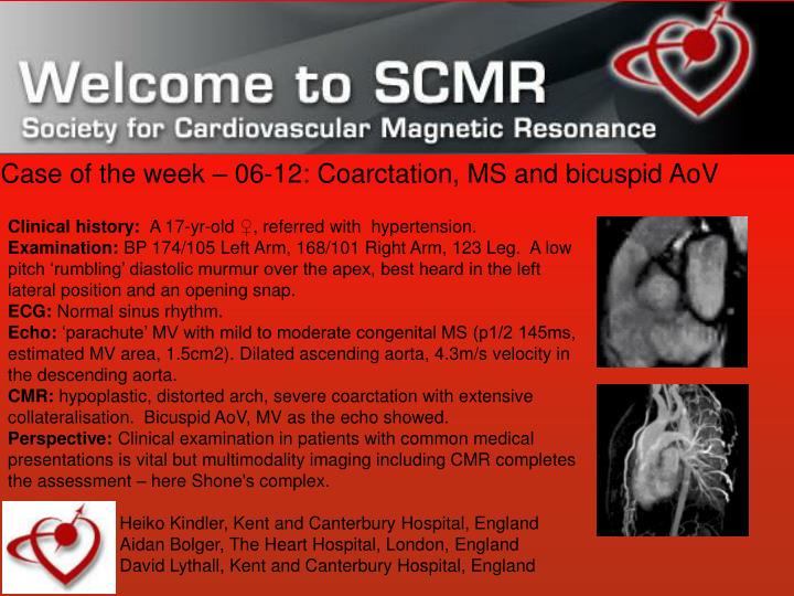
Case of the week – 06-12: Coarctation, MS and bicuspid AoV Clinical history: A 17-yr-old ♀, referred with hypertension. Examination: BP 174/105 Left Arm, 168/101 Right Arm, 123 Leg. A low pitch ‘rumbling’ diastolic murmur over the apex, best heard in the left lateral position and an opening snap. ECG: Normal sinus rhythm. Echo: ‘parachute’ MV with mild to moderate congenital MS (p1/2 145ms, estimated MV area, 1.5cm2). Dilated ascending aorta, 4.3m/s velocity in the descending aorta. CMR: hypoplastic, distorted arch, severe coarctation with extensive collateralisation. Bicuspid AoV, MV as the echo showed. Perspective: Clinical examination in patients with common medical presentations is vital but multimodality imaging including CMR completes the assessment – here Shone's complex. Heiko Kindler, Kent and Canterbury Hospital, England Aidan Bolger, The Heart Hospital, London, England David Lythall, Kent and Canterbury Hospital, England