Download
1 / 64
640 likes | 849 Views
Diagnostic Tests for Cardiovascular System. Diagnostic Tests What you need to know. Client Teaching and Nursing Care Explain procedure to client Know what assessments and interventions nursing may need to do Allergies, Mobility, Nutrition Status Physically, prepare the client.

E N D
Diagnostic Tests What you need to know • Client Teaching and Nursing Care • Explain procedure to client • Know what assessments and interventions nursing may need to do • Allergies, Mobility, Nutrition Status • Physically, prepare the client
DIAGNOSTIC IMAGING • RADIOGRAPH EXAMS • Show heart size, shape, position and outline of shadow • Show lung congestion • Heart failure: pleural effusion from L heart failure • FLOUROSCOPY • An actionpicture radiograph • Allows observation of movement
Chest X-ray • 1 or 2 views (AP and Lateral)
DIAGNOSTIC IMAGING • ANGIOGRAM • A series of radiographs taken after injection with contrast medium • Visualizes: heart, aorta, inferior vena cava, pulmonary artery and vein; coronary arteries • Aids in diagnosing vascular occlusions, pooling in the heart chambers, congenital abnormalities
DIAGNOSTIC IMAGING • ANGIOGRAM (cont.) • Angiography/Cardiac Catheterization/ EP Study • Procedure: involves passage of a catheter through a peripheral vessel heart chamber in order to: • Measure pressure within the heart • Measure blood volume related to cardiac competence • Valvular defects; congenital abnormalities
DIAGNOSTIC IMAGING • Angiogram (cont.) • IV site required • Moderate sedation used, so NPO status for 6-8 hours required. • Intravascular contrast used, all precautions apply • “Recovery Period” after sedation with frequent vital signs • If femoral artery used- bed rest- HOB flat, head on the pillow, movement of affected leg prohibited • Bleeding from puncture site and/or arterial clot are risks • Full day or overnight hospital stay
DIAGNOSTIC IMAGING • Aortogram • Abdominal Aorta + major leg arteries • Viewed after contrast medium administered • Can diagnose aneurysms and other abnormalities
Computed Tomographyaka CT or CAT Scan • X-ray that uses cross-sectional views to image tissue density differences • May use IV contrast to view vascular structures and enhancement of tumors
CT Angiography • 3D reconstruction of CT with contrast Ask about allergies to injectable contrast or shellfish • Used for vascular structures including coronary arteries Radiation exposure is considerable
Cardiac Catheterization • Right Heart Catheterization • Catheter in inserted into a central vein and advanced until enters the Right Atrium, • Pressures are measured in the RA, RV, Pulmonary Artery and Pulmonary Capillaries
Left-heart Catheterization • Left Heart Catheterization • Catheter is inserted into an artery and advance until enters the Left Atrium • Pressures are measured in the LA and LV • A Ventriculogram is done to measure the ejection fraction
Coronary Angiography • During a Left Heart Catheterization, the Coronary Arteries are engaged by the catheter and contrast is injected to determine blood flow through the artery
Electrophysiology Studies(EP Studies) Maps electrical pathways and foci of abnormal heart rhythms • Multiple catheters are inserted in to different veins and sometimes arteries to reach the chambers of the heart and induce dysrhythmias • If the focus of the dysrhythmia is determined, it is then ablated (burned)
MRI/MRA • Soft tissue discrimination • MRI (Magnetic Resonance Imaging) • Looks at structure (Heart defects), function (EF, contractility, relaxation), and areas of scarring in 2D in the heart using Gadolinium contrast • MRA (Magnetic Resonance Angiography) • Looks at the lumen of arteries using GAD to reconstruct vascular structures in 3D
Nuclear Medicine • PET Scan • Viable cardiac tissue will have more glucose (fluorodeoxy) uptake than scarred non-viable areas • Thallium Imaging • Perfusion imaging agent used during exercise or pharmacological stress testing to look for areas of decreased perfusion • Persantine/Adenosine/Dobutamine • Pharmacological stress testing by causing vasodilatation. Perfusion imaging agents Technetium or Thallium. Normal segments will vasodilate more than diseased areas
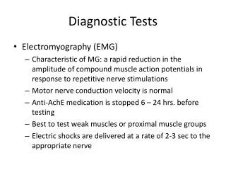
DIAGNOSTIC IMAGING • ELECTROCARDIOGRAPHY • EKG, ECG • A graphic study of the electrical activity of the heart muscle To determine transmission of cardiac impulses through the muscle and conductive tissues
DIAGNOSTIC IMAGING • ELECTROCARDIOGRAPHY cont. • 3 distinct waves or deflections • P Wave • QRS Complex • T Wave • Depolarization = heart contraction • Repolarization = heart relaxation
Electrocardiography (ECG/EKG) • 12 Leads • 12 different angles of the heart can be viewed at once for comparison • Can be used to see • Myocardial ischemia/infarction • Hypertrophy of the myocardium • Dysrhythmias • Drug toxicity • Electrolyte imbalances
Holter Monitoring/ Event Monitoring • A Holter Monitor is an EKG monitor worn by the patient for 24 hours to evaluate for cardiac dysrrhythmias • No Showers! • A journal is kept to match symptoms with dysrrhythmias
Holter Monitor • An Event Monitor may be worn for longer periods of time and has a record button for the patient to push when they feel symptomatic • A Loop Recorder is a monitor which is surgically implanted in the patient to monitor the EKG for longer periods of time
Pacemaker Interrogation • Machine reads pacemaker activity and pacemaker settings to ensure proper timing and strength of electrical impulses • Also used to determine how many “shocks” an internal defibrillator has given and whether or not the “shocks” were appropriate
Echocardiography • Transcutaneous Echocardiography • Probe is used on the skin of the anterior chest to image the heart by ultrasound • Transesophageal Echocardiography (TEE) • Endoscopic probe is inserted into the esophagus and doppler is used to image the heart from the posterior angle, moderate sedation is required
Echo http://depts.washington.edu/anesth/tips/tee_pictures/2009_03_1.gif
Echocardiography • Echo is used to evaluate Left Ventricular function (ejection fraction*), Right Ventricular function, heart valve function, heart wall motion, pericardial sac. *Ejection Fraction is the percentage of Left Ventricular volume the LV ejects with each contraction
Echocardiography • Also used to: • Detect collection of blood or fluid in pericardial sac • Cardiac chamber(s) size and contents • Septum: motion and thickness • Congenital heart disorder • Valve function
Exercise/Stress Test • Pt is asked to exercise using a treadmill or exercise bike, OR • Pt is given a pharmacological agent to induce stress on the heart • EKG and possibly Transcutaneous Echo are monitored as well as vital signs and subjective data from the pt • A positive (symptomatic) stress test will require more evaluation for heart disease
Ankle systolic pressure Higher brachial artery systolic pressure Understanding the ABI The ratio of the higher brachial systolic pressure and the higher ankle systolic pressure for each leg: ABI =
ABI Procedure http://www.nhlbi.nih.gov/health/dci/Diseases/pad/pad_diagnosis.html
Calculate the ABI • For the left side, divide the left ankle pressure by the highest brachial pressure and record the result. • Repeat the steps for the right side. • Record the ABIs and place the results in the medical record. Right Leg ABI Left Leg ABI Right Ankle Pressure Left Ankle Pressure Highest Arm Pressure Highest Arm Pressure ABI Interpretation ≤ 0.90 is diagnostic of peripheral arterial disease Hiatt WR. N Engl J Med. 2001;344:1608-1621; TASC Working Group. J Vasc Surg. 2000;31(1Suppl):S1-S296.
Right ABI 80/160=0.50 Left ABI 120/160=0.75 ABI (Normal >0.90) Highest brachial SBP Brachial SBP 160 mm Hg Brachial SBP 150 mm Hg PT SBP 120 mm Hg PT SBP 40 mm Hg Highest of PT or DP SBP DP SBP 80 mm Hg DP SBP 80 mm Hg Using the ABI: An Example ABI=ankle-brachial index; DP=dorsalis pedis;PT=posterior tibial; SBP=systolic blood pressure.
Interpreting the Ankle-Brachial Index Adapted from Hirsch AT, et al. J Am Coll Cardiol. 2006;47:e1-e192. Figure 6.
Bone Marrow Aspiration • Occasionally pt will take a sedative • Pt will lay prone or on their side • The skin over the bone to be sampled is cleaned with antiseptic • Local anesthetic is then injected into a small area of skin and tissues just over the bone. • A long, large bore needle is used to aspirate samples
Serum (blood) Tests • Cardiac Enzymes • Complete Blood Count (CBC) • Coagulation Studies • Lipids • Metabolic Panel (BMP or CMP) • Electrolytes • Miscellaneous Tests
Serum Cardiac Markers • Proteins released into the blood in large quantities from necrotic heart muscle after an MI. • Include Cardiac Enzymes + Troponin I Important screening diagnostic criteria for an MI
Cardiac Enzymes • Include: • CreatineKinase (CK) • CreatinePhosphokinase (isoenzyme of CK) (CK-MB) • Normal Range: • CPK or CK (Myoglobin) • Normal 26-174 units/L • CK-MB (CreatineKinase, myocardial muscle) • Normal < 5% of total CK • 1.3 to 8.7 units/L
Cardiac Enzymes • Start to rise 2-3 hrs after the beginning of an MI peak in 24hrs return to nml. within 24-40 hrs • Note: CK-MB can be elevated in skeletal muscle damage r/t surgery, trauma, or disease process
Serum Cardiac Markers • Other: Troponin I – a myocardial muscle protein released into circulation after myocardial injury • 2 subtypes: Troponin T and Troponin I • Can identify small amts of myocardial damage