Download
1 / 44
440 likes | 621 Views
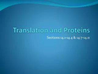
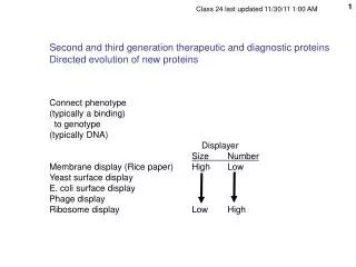
Second and third generation therapeutic and diagnostic proteins Directed evolution of new proteins Connect phenotype (typically a binding) to genotype (typically DNA) Displayer size Number Membrane display (Rice paper) High Low Yeast surface display E. coli surface display

E N D
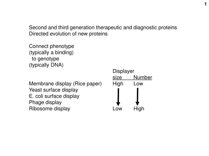
Second and third generation therapeutic and diagnostic proteins Directed evolution of new proteins Connect phenotype (typically a binding) to genotype (typically DNA) Displayersize Number Membrane display (Rice paper) High Low Yeast surface display E. coli surface display Phage display Ribosome display Low High
Screening for natural antibodies using yeast Nature Biotechnology21, 163 - 170 (2003) Flow-cytometric isolation of human antibodies from a nonimmune Saccharomyces cerevisiae surface display library Michael J. Feldhaus, et al. and K. Dane Wittrup C-myc Aga2 becomes associated with Aga1 in the cell. Aga1 becomes covalently anchored to the cell wall c-myc yeast FACS
Av Av Av First purification to get numbers down to < 108 for FACS: magnetic (20 nm) microbeads Avidin-coated magnetic bead μ Bead-labeled cells retained on magnetic column Biotin- B www.miltenyibiotec.com
Created a general scFv library from 58 people using PCR primers from various classes of Igs. • Cloned in E. coli to maintain complexity of 109. • Cloned into yeast for surface expression maintaining complexity • Screen for antigen binding by 2-steps: • Too many cells for initial FACS, so: Magnetic bead capture (avidin micromagnetic beads [20 nm], bind via biotinylated antigen) isolate on column • FACS • Found Abs vs. many antigen tried.: • EGF, EGFR, p53 peptides, fluorescein, etc. • Kd ~ nM range, often. c-myc label Ag label
Isolation of engineered, full-length antibodies from libraries expressed in Escherichia coli. Mazor, Y., Van Blarcom, T., Mabry, R., Iverson, B.L., and Georgiou, G. 2007. Nat Biotechnol25: 563-565. Outer memb., permea-bilized Fortunately, 100 pM avidity, 10x affinity. Divalency? Periplasm Inner memb. Immunize mice spleen cells mRNA PCR VH and VL cDNAs reconstruct full length H&L chains in one plasmid. Clone 107 E. coli with ProteinA/memb anchor fusion protein (“ZZ”) displayed outside inner membrane lysozyme spheroplasts add fluorescent antigen FACS clone E. coli cells, rescue gene Fluorescent antigen ProteinA-E.coli memb. anchor fusion protein cytoplasm Yields regular Abs, no fusion protein.
Recovery of Ag-speific IgGs Off time On time PA = B. anthacis protective antigen
Phage display for a human antibody RT-PCR the rearranged VH and VL cDNAs Reassemble full antibody molecules
Fully Synthetic Human Combinatorial Antibody Libraries (HuCAL) Based on Modular Consensus Frameworks and CDRs Randomized with Trinucleotides Knappik et al, and Andreas Pluckthun and Bernhard Virnekas J. Mol. Biol., 296: 57-86 (2000) CDR3 targeted here VL VH CDRs bkgds colored Framework gray
Starting material here = HuCAL: Fully synthetic human combinatorial antibody library based on modular consensus frameworks and CDRs randomized with trinucleotides. These are single chain antibodies (scFv’s). Framework consensus sequences of 7 of heavy chain V-regions x 7 framework consensuses of light chain V-regions = 49 combinations Plus: randomized CDR3’s by inserting synthetic oligos beween restriction sites. CDR3 = site of VJ (light chain) and VDJ (heavy chain) joinings. CDR = complementarity determining region Knappik et al, JMB, 296: 57-86 (2000)
Found 95% of human Abs are represented by 7 VH and 7 VL exons Constructed: Human Combinatorial Antibody Library (HuCAL) as scFv’s E. coli (2 x 10⁹ recombinants [!]) Made all 49 (7x7) combinations of framework regions [but not represented equally] Mutagenized with triplets to put in all AA substitutions into a run of 6 spots in CDR3 Simulated the AA frequencies found in natural Abs. } cf.
Fully Synthetic Human Combinatorial Antibody Libraries (HuCAL) Based on Modular Consensus Frameworks and CDRs Randomized with Trinucleotides Knappik et al, and Andreas Pluckthun and Bernhard Virnekas J. Mol. Biol., 296: 57-86 (2000) CDR3 targeted here VL VH CDRs bkgds colored Framework gray
Different amino acid combinations achieved after insertion of synthetic oligonucleotides. Planned Found Figure 7. Comparison between design and experimental composition of CDR3 libraries used. For each position of the CDR3 region (numbering according to Kabat et al., 1991; for HCDR3 the position before H101 is numbered 100z, the length variable region is numbered from H95 to H100s), the amino acid composition in the planned libraries (P, left columns) is compared with the composition found from sequencing 257 clones of the initial libraries (F, right columns). The TRIM mixture indicates the mixtures of trinucleotides used in the oligonucleotide synthesis (see Table 3 of the Supplementary Material). Occupied indicates the number of amino acids encoded by the respective mixture and found in the sequenced clones, respectively. AAs There’s a similar table for VL Knappik et al, JMB, 296: 57-86 (2000)
From Biacore: Kd (nM) on time (t1/2) off time (t1/2) * * From subsequent mutagenesis and ribosome diplay
Nature Biotechnology (2000) 18:1287 1 2 6 3 5 4
Start here: Library in DNA form via an E. coli S30 extract Primer must carry T7 promoter Insulin (target)
Ribosome display Absence of a stop codon is necessary to prevent dissociation of the newly synthesized protein from the ribosome. biotin RT-PCRproduct of affinity captured material antigen Affinity selection in rabbit reticulocyte ribosome display is antigen-dependent and requires the absence of a stop codon to keep the protein-ribosome-mRNA complex together. In the presence of a stop codon, the protein synthetic complex dissociates. From Hanes et al., FEBS Letter, 450, 105-110, 1999)
3’ stem-loop Prokaryotic ribosome binding site 5’ stem-loop T7 promoter 3’ AA spacer scFv DNA mRNA DNA construct assembled by ligation and PCR: no cloning in E. coli Here: 2 x 10⁹ molecules. Stem-loops protect frm degradation Later conservative estimate of no. of molecules that can be screened: 2.6 × 10¹¹ per ml of reaction (Nature Methods 4:269 (2007)) scFv: single chain Fragment of variable chain (VH + VL
CDR3 targeted here VL VH CDRs bkgds colored Framework gray
Back to: Nature Biotechnology (2000) 18:1287 Primer must carry T7 promoter 1 2 via an E. coli S30 extract 6 3 5 4 Insulin (target) Anti-insulin? B= biotin B Avidin bead
35S-labeled anti-insulin “Cold” (unlabeled) anti-insulin B B B B B B B B dish Radioimmune assay (RIA) Specific insulin binding activity (gray-white bars) was evident over a smaller amount of non-specific (or different-specific) binding, as assayed by binding of 35S-labeled translated protein in the translational complex. Insulin
Plenty of framework mutations introduced by the PCR (i.e., not in the HuCAL library) Varied in the HuCAL library (CD3) Length variations
7x7 grid Only a 8 of the 49 possible framework combinations were used. Three were used most often. So there may be a preference for particular frameworks for this antigen.
“Consensus WT” HuCAL sequence(without the PCR mutations) All selected clones exhibit tighter binding. That is, the addiitonal mutations introduced by PCR were selected. Entirely cell-free: no bottlenecks due to relatively inefficient DNA transfer steps. One is always dealing here with populations of molecules. Eventually clone in a plasmid in E. coli for expression and testing.
Analogous functions in the same organism; e.g. families of proteases Same function in a different organism; e.g., interferons DNA shuffling (Stemmer - Maxygen) (by PCR) Fragment DNA with DNase Reassemble fragments “recombination” via PCR priming Iterate PCR (no oligo primers) • Base changes can be purposely introduced prior to shuffling • by chemical mutagenesis) • by error-prone PCR during the process • by “faithful” PCR during the process (lower level of mutation) • by using different members of a gene family (paralogs or homologs) Stemmer, W., Proc Natl Acad Sci U S A. 1994 Oct 25;91(22):10747-51.
From: Advances in directed protein evolution by recursive genetic recombination: applications to therapeutic proteins Aaron L Kurtzman et al. Current Opinion in Biotechnology 2001, 12:361–370 • Why alter a natural protein? • For therapeutic proteins: • Tighter target binding (enabling lower doses, perhaps) • Enable desirable side reactions • Disable undesirable side reactions • Improve half-life in bloodstream • Improve production characteristics (e.g., secretion, stability) • For industrial enzymes: • Altered (even new) substrate specificity • Greater stabilty • More robust (e.g., function at extreme pH’s) • Faster
Example 1: Human interferon alpha (IFNa ) 20 IFNa genes in a gene family Each human protein is poor at inducing virus resistance in mouse cells Shuffle the family, Only 2 rounds. Mixing the non-conserved a.a. differences Select for induction of viral resistance in mouse cells Achieve 185X more activity in shuffled protein. Best was even 3.5 X more active than the best mouse IFN. And still equally effective vs. human cells. So an example of a dramatic change in biological specificity (binding?)
Shuffling an engineered human gene with a mouse gene Boxed nts are not conserved. So lots of differences here. Low temp PCR was required due to the poor homology (Klenow instead of Taq) Stemmer, W., Proc Natl Acad Sci U S A. 1994 Oct 25;91(22):10747-51.
Example 2: Antibodies: breaking the natural limit on affinity selection Natural affinity ceiling of 10-10M (100 pM): Kd = off time / on time Endocytosis rate ~~ 10 min to several hours So no selection for off –rate longer than ~3 h (104 sec) Diffusion-limited on-rate ~ 10-6 M/s Selection limit of affinity of natural antibody evolution = off rate / on rate = (1/10-6)/(1/10+4)= 10-10 = Kd --------------------------------------------------------------------------------------------------- J Foote and HN EisenKinetic and Affinity Limits on Antibodies Produced During Immune ResponsesPNAS 1995; 92: 1254-1256
Off time (t1/2) 2x10⁻⁶ per second = 5 days half-life Iterations
Directed evolution of antibody fragments with monovalent femtomolar antigen-binding affinity. Eric T. Boder, Katarina S. Midelfort, and K. Dane Wittrup. Proc Natl Acad Sci U S A. 2000; 97(20): 10701–10705. Used the FACS to selected single chain anti-fluorescein antibodies displayed on the surface of yeast cells. Competed with free fluorescein. DNA shuffled. 4 cycles. Selected for slow off times. Achieved 50 fM affinities. That’s femtomolar, 50 x 10-15M = 5 x 10-14M = 0.05 pM (compare 100 pM above) Slower off-rate than even biotin-avidin (>5d).
Other selections than Ag binding: scMAb + DNA shuffling + ribosome display + selection in DTT (reducing agent) Ab folding without need for disulfide bond (-SH oxidation to –S-S- not needed) as well as high ligand affinity Lutz Jermutus, Annemarie Honegger, Falk Schwesinger, Jozef Hanes, and Andreas Pluckthun, PNAS 98:75-80 (2001)
Example 3: Enzyme stabilization: PAI-1, a protease inhibitor (TPA inhibitor) Error prone DNA shuffling 245X increase in stability (days) Assay = tPA binding Found 11 aa changes, presumably affecting protein folding Back-cross to remove non-contributory mutations: DNA shuffle best clone with original WT DNA. Maintain selective pressure. Analyze “progeny”: see 2 of the 11 aa changes lost, not needed, replaced by WT sequence. J Mol Biol. 2001 Jan 26;305(4):773-83. Different structural requirements for plasminogen activator inhibitor 1 (PAI-1) during latency transition and proteinase inhibition as evidenced by phage-displayed hypermutated PAI-1 libraries. Stoop AA, Eldering E, Dafforn TR, Read RJ, Pannekoek H.
Example 4: Viral tropism: murine leukemia virus, a retrovirus • MLV, 6 strains, all poor infection of CHO cells. • DNA shuffled envelope gene of 6 strains • chimeric virus that can infect CHO cells And selected incidentally for non-inactivation under conditions of laboratory manipulation (100X centrifugation resistant) Nat Genet. 2000 Aug;25(4):436-9. Molecular breeding of viruses. Soong NW, Nomura L, Pekrun K, Reed M, Sheppard L, Dawes G, Stemmer WP.
A more global version of DNA -- genome shuffling: A different (unnatural) kind of genetic mixing using whole genomes Zhang et al., Nature 415, 644 (2002) Can mix Streptomyces (bacteria) genomes by protoplast fusion. effective diploid bacteria The fused cells will generate recombinant haploid spores. Target – tylosin production (an antibiotic) Mutagenize a culture, collect 22,000 survivors. Screen them all for tylosin synthesis, pick the top 11. Protoplast fuse all 11 with each other. Collect 1000 progeny. Screen 100 for tylosin, collect the best 7. Protoplast fuse again. Collect 1000 again. Screen for tylosin again. Characterize the best 2: Tylosin production is up 9-fold. So productivity is up 9-fold, without a tremendous amount of work (22000 screen the most)
A more supervised version of DNA shuffling Multivalent avimer proteins evolved by “exon” shuffling of a family of human receptor domains Nature Biotechnology 23: 1556 (2005) Joshua Silverman, et al & Willem Pim C Stemmer Avidia, Inc A misnomer; really domain shuffling) Strategy: Create therapeutic proteins by combining hundreds of known binding domains from receptor proteins in new random combinations and selecting for binding to a specific target by phage display
Organization of binding domains in typical mammalian receptors A-domains:(~35-40 AA’s/domain): determine binding speficicity of many receptors Typical receptor structures of 217 A-domains (~metaphorically?) as a spacer between domains Dual specificity domain Bipartite domain 2 domains cooperating Degenerate oligos synthesized to coding for 35-40 AAs of the A domains Only AA’s naturally found at each position were coded for. Conserved structural AAs were kept constant. Complexity = 1023 .Actually realized = 1010 as phage display particles Select one domain at a time, serially, by panning: LRP = LDL receptor related protein
Isolation of a high affinity binding protein to IL6 ( interleukin 6 ) by iterative selection (IL6 is a target for cancer and inflammation) Phage display (M13) IL6 immobilized on plates. Recovered proteins from first cycle, cloned and tested for IL6 binding; 20 top binders pursued. Add the domain library to each of the 20 first round winning domains. Again pick best 20 overall. After a third cycle pick the very best binder: = “C326” Monomer protein Screened for binding Monomer displayed on phage coat Build 20 dimers pools from 20 best monomers One domain Three domains Two domains M13 phage IL6 = interleukin 6
Add an IgG-binding domain at the end to prevent rapid clearance (measured half-life of 89 hours in monkeys)
Binding measured by a competition assay (“AlphaScreen”) Reactive oxygen species can react only over a short distance with and “acceptor” bead Laser Luminescence Reactive Oxygen IL6 receptor IL6 Avidin bead: biotinylated IL6 : gp130-Fc : Protein A bead Competition: IL6 (non-biotinylated) or C326 avimer
Physical assay: Biacore surface plasmon resonance to measure binding kinetics
Biological assay: Stimulation of proliferation of TF-1 cells (erythroleukemia line) 16 h of 3H-TdR incorporation to measure promotion of DNA synthesis Commercial anti-IL6 antibodies
Acute phase inflammatory response induced by IL6 is reversed by avimer C326 (in mice) Specific for IL6-induced inflammation