Download
1 / 47
480 likes | 485 Views
MCB100 Introductory Microbiology 19 Sept. 2018 - Chapter 4 Microscopy. MCB100 Exam 1 Fall 2018 Friday, September 28, 2018 2:00 – 2:50 pm Covers: chapters 1, 2, 4, 3 & 6 (Chapter 5 is not on exam 1!)

E N D
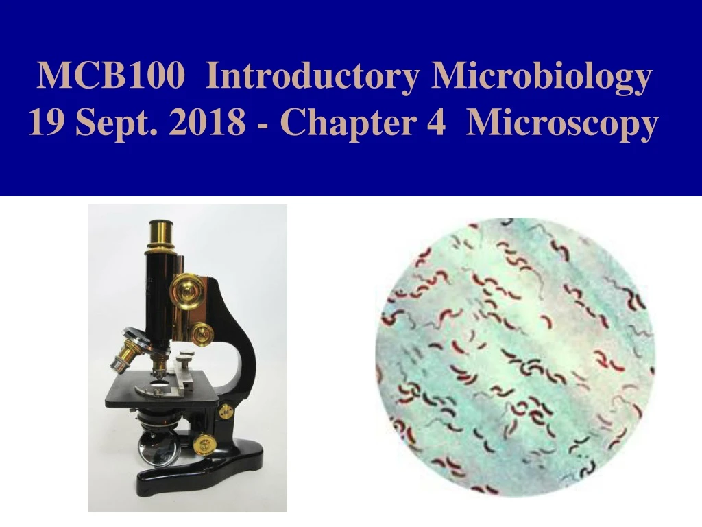
MCB100 Introductory Microbiology 19 Sept. 2018 - Chapter 4 Microscopy
MCB100 Exam 1 Fall 2018 Friday, September 28, 2018 2:00 – 2:50 pm Covers: chapters 1, 2, 4, 3 & 6 (Chapter 5 is not on exam 1!) Place: If your last name starts with A – M: please take the exam in room 2079 NHB If your last name starts with N – Z: please take the exam in room 100 Noyes Lab Review Session: 7:00 – 7:50 pm, Wednesday, Sept. 26 Place: Room 161 Noyes Lab
MCB100 Exam 1 Fall 2018 Practice Exam 1 has been updated. To find the link to the practice exam, go to:www.life.illinois.edu/mcb/100 - Click on: “Exam Information” - Scroll down to find the link to the practice exam. (It downloads as a MS Word Document.)
Do you have a conflict for Exam 1, which is scheduled for 2 – 2:50 pm on Friday 9/28/2018? Contact Dr. Chapman by e-mail: kenchap.life.illinois.edu What is the nature of your conflict? The conflict exam will be given at 3 pm on Thursday, 9/27/18 in room 242 Burrill Hall. Does that fit into your schedule? (It's possible to do it earlier in the day but it's more difficult to find a quiet room where a student can take an exam when the lab is being used by a class.)Conflict exams must be taken before the rest of the class takes the test. Sick on the day of the exam? Don’t come and fail the test because you feel so bad you can’t think. See a doctor and take care of your health. Also get an excuse note. There are no make-up exams in MCB100, but in the event of illness or unexpected circumstances your score can be prorated. If you take a test, your score will be counted as it is. You can’t take a test and later decide you’d rather have your score prorated.
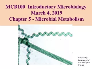
Anthony Van Leeuwenhoek used a home-made simple microscope to make the first reported observations of bacteria and other microbes in the late 1600s.
Late 1600s Robert Hooke used compound microscopes to examine small plant and animal specimens. He observed the compartmentalized structure of cork and introduced the use of the term “cell” in biology.
Development of Microscopes Which one the following events in the development of microscopy occurred first? A. first photograph of bacteria was taken B. the compound optical microscope was invented C. the glass lens was invented D. first visual observation of bacteria E. first visual observation of a virus
Magnification: the ability of a microscope or a lens to increase the apparent size of the image of an object. Total Magnification: For a compound microscope the total magnification is the product of the magnifying abilities of all of the lenses in the system. Magnification Magnification Total Magnification = of the x of the Objective Lens Ocular Lens
Resolution: The ability to discern fine details in an image of an object. Resolving Power: (AKA: Resolution Distance) R.P. is a measure of the smallest distance between two separate objects that can be seen to be two separate objects. The Resolving Power Formula R.P. = 0.61 x wavelength___ numerical aperture Smaller R.P. = ability to see finer details To get better resolution (a smaller R.P.) you need to increase the numerical aperture or decrease the wavelength of the light used to illuminate the object.
Total Magnification of a Compound Microscope If the magnifying power of the objective lens is 90x and the magnifying power of the ocular lens is 11x, what is the total magnification? A. 79x B. 101x C. 990x D. 1199x E. 9011x
The Limits of Resolution with Light Microscopy Typical bacterial cells are 700 – 3000 nm in length. Typical viruses are smaller than 100 nm in diameter. For light with a wavelength of 480 nm (blue) using a good microscope with a numerical aperature of 1.25… Resolving Power = 0.61 x 480 nm / 1.25 = 235 nm - Viruses are too small to see with a light microscope and bacteria are so small you can’t see much detail.
The electron microscope allows you to see viruses and subcellular structures such as ribosomes, because electrons have a shorter wavelength than light and thus smaller Resolving Power (better). With a better Resolving Power you can use higher magnifications without encountering the phenomena of “empty magnification”. With a good light microscope the greatest practical magnification is about 1000x. Electron microscopes can be used at up to 100,000x magnification.
Microscopy Techniques A. Bright-field Microscopy B. Dark-field Microscopy C. Phase Contrast Microscopy D. Fluorescence Microscopy E. Electron Microscopy
Bright-field Microscopy Light shines through the specimen and directly into the objective lens and to the observer’s eye creating a lighted or bright field of vision. Bacteria and other microorganisms are seen as dark objects on a white background. This works best when the bacteria are stained a bright color and immobilized. Bright-field microscopy has the advantage of being easy to use. The disadvantage is that it works best with fixed and stained microorganisms (dead). Dark-field Microscopy Light shines on the specimen from an oblique angle and does not go directly into the objective lens. The background or field of vision is dark. The only light that gets to the observer’s eye is the light that is scattered by the specimen. The specimen does not have to be stained. This technique works for live microorganisms but they move around pretty fast making it difficult to focus on them.
Dark-Field Microscopy Figure 4.7
Microscopy 1 Choose the FALSE statement about microscopy. A. One can’t see viruses with a light microscope even if the objective lens is of excellent quality. B. If you want to see unstained bacteria darkfield microscopy is better than brightfield microscopy. C. The compound microscope was invented by Anthony Van Leeuwenhoek. D. The Gram stain was invented in 1884.
Phase Contrast Microscopy Invented in 1934 by Frits Zernike, phase contrast microscopy relies on a complicated optical trick to enhance the visual effect seen when light goes from a medium of lesser optical density (water) to a medium of greater optical density (cytoplasm). Beams of light are bent when they cross the boundary between two different transparent substances with different optical densities. A lens can diffract a beam because light moves slower in glass than in air.
The bending of light at the boundary of two phases allows you to see the phase boundary even if both phases are transparent. In phase contrast microscopy the optics enhance the contrast seen when light is diffracted at a phase boundary.
Phase Contrast Microscopy is most useful for examining living protozoa and other eukaryotic cells. It does not require the specimen to be stained. Epithelial cell in bright field microscopy vs. phase contrast.Source: wikipedia
Fluorescent Microscopy In Fluorescent Microscopy the specimen is illuminated with UV light which can cause some chemicals to emit visible light. UV light is called black light because you can’t see it. The only visible light seen is that which is produced by fluorescence of naturally occurring compounds in the microorganism or special dyes that are used to stain the specimen.
Natural fluorescence - Some bacteria (ex: Pseudomonas aeruginosa) make a fluorescent pigment - Photosynthetic pigments like chlorophyll are fluorescent - Methanogenic archaea make a fluorescent pigment Fluorescent dyes for DNA (acridine orange) for Bacillus anthracis (fluorescein isothiocyanate) for Mycobacterium tuberculosis (auramine O) Immunofluorescence microscopy A fluorescent dye such as fluorescein (yellow-green) or rhodamine (red) is chemically joined to an antibody to make a very specific probe. The green algae Chlamydomonas stained with 2 fluorescent antibodies.Merchant et al. in Science 10/12/2007
Fluorescent Micrographs of Cyanobacterial Cells Green color is from fusion of GFP to the FtsH gene. Red is from natural fluorescence of photosynthetic pigments. Merged image shows non-photosynthetic cells.from: Conrad Mullineaux, University of London
Microscopic Techniques What type of microscopy would be the most likely to be used by a pathologist who wants to examine a tissue sample for the presence of abnormal-looking (cancerous) cells? A. Dark Field Microscopy B. Bright Field Microscopy C. Phase Contrast Microscopy D. Immunofluorescence Microscopy
Electron Microscopy General features: - The sample is bombarded with electrons - The image is produced using a cathode ray tube - Sample must be dehydrated - Sample must be stained with a heavy metal - Sample must be in an evacuated chamber TEM - Transmission Electron Microscopy - thin sliced sample - used to see cellular organelles such as E.R., mitochondria, chloroplasts, ribosomes etc. SEM - Scanning Electron Microscopy - sample is sputter coated with gold - used to look at the surface of objects (variant = freeze-fracture etching)
Electron Microscopy General features: - The sample is bombarded with electrons - The image is produced using a cathode ray tube - Sample must be dehydrated - Sample must be stained with a heavy metal - Sample must be in an evacuated chamber TEM - Transmission Electron Microscopy - thin sliced sample - used to see cellular organelles such as E.R., mitochondria, chloroplasts, ribosomes etc. SEM - Scanning Electron Microscopy - sample is sputter coated with gold - used to look at the surface of objects (variant = freeze-fracture etching)
Stains for Light Microscopy Simple Stains Basic dyes are used to stain the microorganisms. The chromaphore has a positive charge & binds to negatively charged groups on the bacterial cell wall. Negative StainsAcidic dyes are used to stain the area around the microorganism. The chromaphore has a negative charge and sticks to glass. Differential Stains Two or more dyes are used and different microorganisms stain different colors. This is caused by differences in the structure or chemical make-up of the cell surface of the microorganisms. Examples of Differential Stains - The Gram Stain - Schaeffer-Fulton Endospore Stain - Ziehl-Neelsen Acid-Fast Stain
Simple Stains are used to stain microorganisms a bright color to make them more visible in Bright-Field Microscopy. Dyes used for simple stains are called basic dyes or cationic dyes. Basic dyes are fairly complicated organic molecules that ionize in water. When a compound ionizes it breaks down to an anion and a cation. For a basic dye the chromophore (colored portion) is a proton acceptor. This makes the chromophore positively charged in aqueous solution. Many components of the bacterial cell wall and cytoplasmic membrane are acidic and will be deprotonated in water, this makes the cell envelope negatively charged. Examples of Basic Dyes Crystal Violet Safranin Methylene Blue
Negative Stains – Acidic Dyes For an acidic dye the chromophore is a proton donor when the dye molecule is dissolved in water. This makes the colored reagent negatively charged. Acidic dyes usually don’t stain microorganisms but they can stain glass. The negative stain is a procedure that is used to visualize the bacterial capsule. A capsule is a slimy layer of polysaccharides that coats the outside surface of some bacteria and protects the bacterium from the host immune system. Acidic Dyes Nigrosin India Ink Congo Red Eosin
Differential Stains - The Gram Stain - Schaeffer-Fulton Endospore Stain - Ziehl-Neelsen Acid-Fast Stain A differential stain uses two different dyes to distinguish between different types of cells or different parts of cells. The first dye used is called the primary dye. The primary dye is generally followed by some sort of wash. Then the second dye or counterstain is applied.
The Gram Stain The Gram stain is used to divide bacteria into two groups based on the thickness of the cell wall. Bacteria with thick cell wallswill appear purple and are called Gram positive. Bacteria with thin cell walls will appear pink and are called Gram negative. Examples of Gram Positive Bacteria: Clostridium tetani, Streptococcus pneumonia, Corynebacterium diphtheria Examples of Gram Negative Bacteria: Esherichia coli, Salmonella sp. Vibrio cholera, Yersinia Pestis
Ziehl-Neelsen Acid-Fast Stain The Acid-Fast stain is used to identify bacteria in the Genera Mycobacteria and Nocardia, including the clinically very significant bacterium Mycobacterium tuberculosis. The basis of this differential stain is that the cell walls of the Mycobacteria contain a lot of hydrophobic waxy mycolic acids. The dye Carbol Fuchsin sticks tightly to the waxy mycolic acids. Decolorization with acidified alcohol removes the Carbol Fuchsin from other types of bacteria. The counterstain is typically methylene blue.
Schaeffer-Fulton Endospore Stain The Endospore Stain is used to identify spore forming bacteria in the Grampositive genera Bacillus and Clostridium. It is especially useful for identifying: Clostridium botulinum, Clostridium tetani, C. perfringens & Bacillus anthracis. The endospore cell wall is mineralized with dipicolinic acid and calcium. The endospore can be stained by the dye Malachite Green. Most bacterial cells don’t stain well with Malachite Green. The red dye Safranin is used as a counterstain to see the vegetative (non-spore) cells. Endospore Stain Vegetative cells (actively growing)stain red. Spores are green.
Stains for Microscopy What is the purpose of the Ziehl-Neelson Acid Fast stain? A. helps to identify Clostridium andBacillus species B. helps one to detect the presence of a capsule around bacterial cells C. separates bacteria into two groups based on thickness of the cell wall D. helps to identify Mycobacterium species like tuberculosis bacteria