Download
1 / 54
560 likes | 583 Views
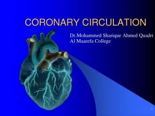
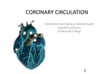
Coronary circulation. Anatomic considerations. Normal coronary blood flow. The resting coronary blood flow= 225 ml/min In strenuous exercise = increase three to four folds. Phasic changes in coronary blood flow. Epicardial Vs. subendocardial blood flow. Control of coronary blood flow.

E N D
Normal coronary blood flow • The resting coronary blood flow= 225 ml/min • In strenuous exercise = increase three to four folds.
Control of coronary blood flow Metabolic regulation Nervous control
Metabolic regulation • Blood flow through coronary system is regulated almost entirely by local arterial vasodilatation in response to cardiac muscle need for nutrients. Increased contraction Increase in rate of coronary blood flow
Oxygen demand. • Vasodilator substances: • Adenosine. • Potassium ions. • Hydrogen ions. • Carbon dioxide. • Bradykinin. • Prostaglandins.
Nervous control • Direct effect: • Direct action of Ach and Nepi on coronary vessels. Sympathetic transmitter Vascular dilatation β receptors Vascular constriction α receptors
Indirect effect: • Symp stimulation HR & contractility Rate of metabolism. • activity local blood flow regulatory mechanisms blood flow increases.
Ischemia: • Lack of oxygen due to inadequate perfusion of the myocardium, which causes imbalance between oxygen supply and demand.
Coronary atherosclerosis • The most common cause of myocardial ischemia. • Epicardial coronary arteries are the major site. • Major risk factors: • Increase in LDL. • Decrease in HDL. • Cigarette smoking. • Hypertension. • DM.
Normal function of vascular endothelium Loss of these defenses • Local control of vascular tone. • Maintenance of an anticoagulant surface. • Defense against inflammatory cells. • Inappropriate constriction. • Luminal clot formation. • Abnormal interactions with blood monocytes & platelets.
Acute coronary occlusion • Thrombosis. • Embolism. • Local spasm: • Direct irritation of the smooth muscle. • Nervous reflexes.
Location of the obstruction • Influence the quantity of myocardial ischemia. • Determines the severity of the clinical manifestations.
Collateral circulation With sudden occlusion. With gradual developing atherosclerosis.
Effects of ischemia • Disturbances of myocardial functions: • Mechanical function. • Biochemical function. • Cell membrane function. • Electrical function.
Effect of ischemia 1)Mechanical function: • Failure of normal muscle contraction & relaxation. • Ischemia of large portions of ventricle : left ventricular failure. • Regional disturbances: • Systolic stretch.
Effect of ischemia 2) Biochemical function: • Fatty acid can’t be oxidized. • Glucose is broken down to lactate. • Reduced intracellular PH and ATP stores.
Effect of ischemia 3) Cell membrane function: • Leakage of potassium and uptake of sodium by myocytes. 4) Electrical function: • ECG changes: • Repolarization abnormalities. • Transient ST segment depression. • Electrical instability: • Ventricular tachycardia and fibrillation.
Ischemic heart disease Stable angina (chronic artery disease) • Acute coronary syndrome: • Unstable angina. • Acute MI.
Stable angina… • An effort-related chest discomfort. • Characteristics: • Heaviness. • Pressure. • Squeezing. • Smothering. • Choking pain.
Stable angina… • Causes: • CAD. • Other heart diseases: • Aortic valve disease. • Hypertrophic cardiomyopathy.
Stable angina… • History: • A man > 50 years. • A woman > 60 years. • Pain with physical & emotional exertion. • Last to 5-10 min.
Stable angina… • Radiating pain to the left shoulder, both arms, back, interscapular region, root of the neck, jaw and teeth.
Stable angina… • physical examination: • Atherosclerotic disease at other sites. • Important risk factors: • Hyperlipidemia • DM. • Left ventricular dysfunction. • Conditions that may exacerbate angina: • Anemia. • Thyroid disease.
Stable angina… • Laboratory examination: • Urine analysis ( DM and renal disease). • Full blood count. • Measurements of: • lipids,. • Glucose. • Createnine. • Hematocrite. • Thyroid function test.
Stable angina… • Other investigations: • Resting ECG: most important baseline investigation. • Stress testing.
Stable angina… • Other investigations: • Coronary arteriography.
Stable angina… • Management: • A careful assessment. • Identification and control of aggravating conditions. • Identifications of high risk pts. • Application of treatment to improve life expectancy.
Stable angina… • Drug therapy: • nitrates. • β-adrenergic blockers. • Calcium antagonist. • Antiplatelet drugs.
Unstable angina… • Angina pectoris that is rapidly worsening. • Characteristics: • Occurs at rest, usually lasting >10 min. • Sever and of new onset. • Crescendo pattern.
Unstable angina… • Causes: • Shares common pathophysiological mechanisms with acute MI. • Plaque rapture or erosion. • Dynamic obstruction ( coronary spasm). • Rapidly advancing coronary atherosclerosis.
Unstable angina… • History: • History of chronic stable angina. • May present as new phenomena. • Chest pain ( substernal region, radiating to the neck, left shoulder and left arm).
Unstable angina… • Physical examination: • Diaphoresis. • Pale cool skin. • Sinus tachycardia. • 3rd or 4th heart sound. • Biochemical markers: • Troponin I & T. • CK.
Unstable angina… • ECG changes: • 12 lead ECG is mandatory. • ST elevation or depression.
Unstable angina… • Management: • Urgent admission to hospital. • Bed rest. • Antiplatelet. • β-blockers (atenolol). • IV or buccal nitrates. • Revascularization.
Stableangina Unstable angina • Fixed stenosis. • Demand-led ischemia. • Predictable. • Exercise tolerance test. • Dynamic stenosis. • Supply-led ischemia. • Unpredictable. • Clinical features. • ECG changes. • Biochemical markers.
Myocardial infarction • Occurs when there are zero flow or so little flow that it can’t sustain cardiac muscle function. • Occlusive thrombus in a coronary artery.
Myocardial infarction • Clinical features: • Pain (sever, last longer). • Breathlessness. • Vomiting. • Collapse. • Syncope.
Myocardial infarction • Investigations: • ECG: • Partial thickness infarctionST/T wave changes. • TransmuralinfarctionST elevation and Q waves. • Biochemical markers. • Chest radiography. • Cardiac US.
Myocardial infarction • Management: • Immediate access to hospital. • High-flow oxygen. • ECG monitoring. • I.V analgesia and antiemetic. • Detect and manage acute complications: • Arrhythmia. • Ischemia. • Heart failure.
Myocardial infarction • Complications of infarction: • Arrhythmia. • Ischemia. • Acute circulatory failure. • Pericarditis. • Embolism.