Download
1 / 23
250 likes | 666 Views
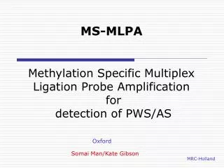
Multiplex Ligation-dependant Probe Amplification. MLPA Tissue Service Sonesh Joshi Kennedy Galton Centre North West Thames Regional Genetics Service Northwick Park & St. Marks Hospital. Introduction. Tissue Service: What we do Analyse chromosomes for aneuploidy

E N D
Multiplex Ligation-dependant Probe Amplification MLPA Tissue ServiceSonesh Joshi Kennedy Galton Centre North West Thames Regional Genetics Service Northwick Park & St. Marks Hospital
Introduction • Tissue Service: • What we do • Analyse chromosomes for aneuploidy • Referrals: Miscarriage, IUD, Stillbirth, TOP • Karyotyping of cultured tissue samples • Typical sample: Villus, skin, cord • Culturing is a long process, needs fresh tissue – Otherwise may not grow • Lots of consumables used • Now by MLPA (service introduced in April 2009) • No culturing required • Uses extracted DNA
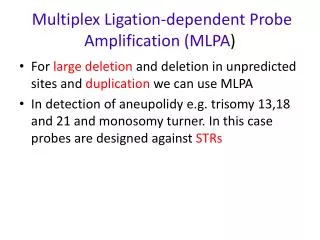
Introduction • What is MLPA? • A multiplex-PCR based method determining copy number change of targeted chromosome regions • Looking at the sub-telomeric regions
Advantages • Tested in batches • Increases efficiency and reduces costs • Reduces reporting time because no culturing required • Use samples that were unable to grow in culture • Increases success rates (major advantage) • MLPA success rate >90% compared to ~70% for karyotyping
Limitations • Unable to detect certain abnormalities • Imbalance in regions not covered by probes • Only sub-telomeres tested • Balanced rearrangements • Test only detects copy number gain or loss • Triploidy • Data normalization prevents whole genome gain being detected, only relative gains and losses • Mosaicism • Only tells you if abnormal or not, due to statistical cut-offs
Quality Data • Abnormality rate is lower due to MLPA being unable to detect certain abnormalities
How is MLPA done? • Tissue Processing • Placental tissue (villus), Skin, Other fetal tissue with DNA • DNA Extraction • Looking at purity of DNA • Contaminated DNA • Inhibit PCR stage of experiment • May give poor results
Hybridisation Sequence PCR Primer Y Stuffer Sequence PCR Primer X Target Sequence How is MLPA done? • MLPA kits used for subtelomeric regions • PO36 and PO70 kits, have 1 probe for each sub-telomere, plus control sequences • One MLPA probe has 2 oligonucleotides From www.mlpa.com
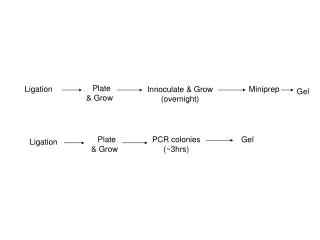
How is MLPA done? • Denaturation and Hybridisation • Sample DNA is denatured and Probe Mix is then added • The 2 probes hybridise to the target sequence (sample DNA) adjacent to each other • Ligation • Hybridised probes are ligated using thermostable ligase From MLPA.com
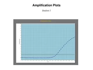
How is MLPA done • PCR • Only ligated probes are amplified From www.mlpa.com • PCR products are size separated by capillary electrophoresis • Using eg: ABI3130 • Relative amount of each probe measured via fluorescence • Determines copy number of each probe • Sample copy number compared to normal control
Normal Fluorescent Amount How is MLPA done • ABI Electrogram • Difference in target probe sequence amount results in different relative peak heights Male or Female X/Yp 1p Size Standard
How is MLPA done • Coffalyser • Statistical analysis software • Compares amount of probe present between sample and controls to provide sample:control ratio for each probe (eg: normal sample:control, all ratio’s should be 1.0) • Blue → Normal • Green → Gain (≥1.3) • Orange → Loss (≤0.7) • White → Ambiguous (Close to the cut off)
How is MLPA done • Relative loss of Xp and Xq sub-telomeres: • Consistent with Turner Syndrome
How is MLPA done • Gain of chromosome 21p and q sub-telomeres: • Consistent with trisomy 21 (Down Syndrome) • All abnormal results are confirmed using a second test (with other kit)
Blank Troubleshooting
Normal Large Q-fragments
Normal Shoulder Peaks
Normal Sloping Peaks
Normal Poor Profile
Poor Profile Troubleshooting • Poor Profile – Possible PCR failure of certain probes • Poor PCR • Incomplete hybridisation due to evaporation • Contaminated DNA • – Or use less DNA when possible to reduce contaminants • Re-do MLPA • Re-precipitate DNA
Acknowledgments • I’d like to thank everyone at the Kennedy Galton Centre for their support • Special thanks to Matthew Edwards and Richard Ellis for their input and advice. • Further Information www.mlpa.com