Download
1 / 72
730 likes | 746 Views
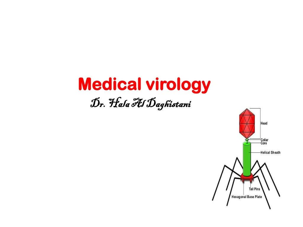
Medical virology. Medical virology Dr. Hala Al Daghistani.

E N D
Medical virology Medical virology Dr. Hala Al Daghistani
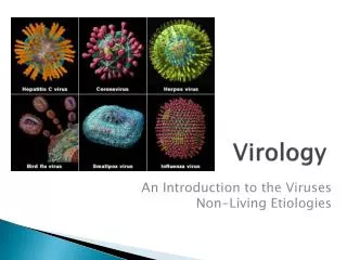
VIRUSES are non-living agents that infect all life forms (plants, phages, and animal viruses). Cultivation needs (tissue culture); they are obligate intracellular parasites). No virus is able to produce its own energy (ATP) to drive macromolecular synthesis.Composition: Contain DNA or RNAContain a protein coat = Capsid that made up of Capsomeres (NA + capsid that formed neucleocapsid). Some are enclosed by an Envelope(enveloped virus), other not (naked virus)Some viruses have Spikes (COH/protein)Most viruses are tissue specific.Only EM allowed for visualization of virusesSpecificTissue tropism is the cells and tissues of a host which support growth of a particular virus . Some bacteria and viruses have a Broad tissue tropism and can infect many types of cells and tissues. Structural Classes1. Icosahedral symmetry2. Helical symmetry3. Non enveloped (“naked”)4. Enveloped
Satillite virus: Viruses that depend on Co-infection (simultaneously) with a helper virus orsuperinfection (carrier, chronic), e.g. Hepatitis delta virus (HDV), cannot propagate without HBV Viroids :Unencapsidated, small circular ssRNA molecules that replicate autonomously, present only in plants, e.g. potato spindle tuber viroid Prions: No nucleic acid, Infectious protein e.g. BSE (bovine spongiform encephalopathy or mad cow disease) Virons (virus): A complete viral particle, consisting of RNA or DNA surrounded by a protein shell and constituting the infective form of a virus.
Principle of viral infections • All viruses package their genomes inside a particle that mediates transmission of the viral genome from host to host • The viral genome contains the informations for initiating and completing an infectious cycle within a susceptible, permissive cell. • Aninfectious cycle includes attachment, and entry of the particle, uncoating, transcription and or translation of viral genome, replication, and assembly and release of particles containing the genome
Attachment(Adsorption) • Virus encounters susceptible host cells • Adsorbs specifically to receptor sites on the cell membrane • Because of the exact fit required, viruses have a limited host range
Penetration • Flexible cell membrane of the host is penetrated by the Whole virus or its Nucleic acid • Endocytosis: entire virus engulfed by the cell and enclosed in a vacuole or vesicle • The viral envelope can also directly fuse with the host cell membrane Uncoating • Enzymes in the vacuole dissolve the envelope and capsid, then virus is uncoated Synthesis and Assembly • Free viral nucleic acid exerts control over the host’s synthetic and metabolic machinery • DNA viruses- enter host cell’s nucleus where they are replicated and assembled • DNA enters the nucleus and is transcribed into RNA • The RNA becomes a message for synthesizing viral proteins (translation) • New DNA is synthesized using host nucleotides • RNA viruses- replicated and assembled in the cytoplasm Releases • Nonenveloped and complex viruses are released when the cell lyses or ruptures • Enveloped viruses are liberated by budding or exocytosis • Anywhere from 3,000 to 100,000 virions may be released, depending on the virus • Entire length of cycle- taks from 8 to 36 hours
How are viruses named Naming based on 1- the disease they cause: poliovirus, rabies virus 2- the type of disease: murine leukemia virus 3- geographic locations: Sendai virus, Coxsackie virus 4- their discovers: Epstein-Barr virus 5- how they were originally thought to be contracted: dengue virus (“evil spirit of Babylonia”) cramp-like seizure, influenza virus (the “influence” of bad air) 6- combinations of the above: Rous Sarcoma virus Taxonomy of viruses • No evidence for common viral ancestor. • Taxonomy from Order downward (3 orders now recognized) • Classification based on type of NA, strategy for replication, and morphology. • Familynames end in –viridae • Many families have subfamilies. Ends in -virinae. • Genusand species names end in -virus. Viral species: A group of viruses sharing the same genetic information and ecological niche (host). Common names are used for species. Subspecies are designated by a number.
The Baltimore classification system Based on genetic contents and replication strategies of viruses. According to the Baltimore classification, viruses are divided into the following seven classes: 1. dsDNA viruses 2. ssDNA viruses 3. dsRNA viruses 4. (+) sense ssRNA viruses (codes directly for protein) 5. (-) sense ssRNA viruses 6. RNA reverse transcribing viruses(ssRNA is reverse-transcribed into ssDNA, this strand of DNA is known as cDNA which is replicated to dsDNA) 7. DNA reverse transcribing viruses. ( dsDNA is transcribed to ssRNA then to mRNA) where "ds" represents "double strand" and "ss" denotes "single strand". All viruses must produce mRNA, or (+) sense RNA . The Baltimore classification has + RNA as its central point - Most genomes are non-segmented (the genome is all on one piece of RNA or DNA).- Some genomes are segmented, meaning there are several fragments of genetic material that make a complete virus genome. - Also, some genomes are linear, meaning that there is a beginning and end to the genome, while other genomes are circular (no beginning and no end|).
Isolation, Cultivation, and Identification of Viruses Viruses must be grown in living cells Bacteriophages form plaques on a lawn of bacteria Animal viruses may be grown in • Cell culture • Embryonated eggs • Living laboratory animals Viral Identification • Cytopathic effects • Serological tests • Identify antibodies against viruses • Use antibodies to identify viruses by neutralization tests, viral hemagglutination • Nucleic acids, PCR
Using Cell (Tissue) Culture Techniques Most viruses are propagated in some sort of cell culture The cultures must be developed and maintained Animal cell cultures are grown in sterile chambers with special media Cultured cells grow in the form of a monolayer
Bacteriophage • Attachment to cell surface receptors - no active movement) • Penetration – only genome enters • Biosynthesis –Production of phage DNA and proteins • Maturation – assembly to form intact phage • Release due to phage induced lysozyme production Results of Multiplication of Bacteriophages Lytic cycle • Lytic or virulent phage • Phage causes lysis and death of host cell Lysogenic cycle • Lysogenic or temperate phage • Phage DNA incorporated in host DNA Prophage
Viral Pathogenesis: Elements of Virus-Host Interaction • Viral strain • Inoculum size • Route of exposure • Susceptibility of host • Is there pre-existent immunity from past exposure or vaccination? • Host genetic factors • Immune status and age of host Steps in Human Viral Infection • Virus may enter through skin, mucous membranes, respiratory tract, GI tract, via transfusion, needle-stick, or maternal-fetal transmission • Local replication at site of inoculation • Neurotropic agents may travel along nerve routes or reach CNS by viremic spread
Pathogenesis of Human Viral Infection • For many agents, there is replication in regional lymph nodes with subsequent viremia and spread to target organs: e.g. Some travel free in plasma (e.g. picornaviruses); some are cell associated (e.g. cytomegalovirus) • Non-specific and virus-specific host immune responses come into play to downregulate viral replication Immune Response to Viral Infections • Non-specific immunity • Phagocytic cells (neutrophils and monocyte-macrophages) • Cytokines (e.g., interferons) • Natural killer cells • Other ‘antiviral’ factors • Specific immunity • Antigen specific B and T cell responses • Antibodies • Cytotoxic T cells • ADCC
The Effect of Virus Infection on the Host Cell Cytopathic Effects 2. Inhibition of Host Macromolecular Biosynthesis 3. Changes in the Regulation of Gene Expression 4. Appearance of New Antigenic Determinants on the Cell Surface (If it is enveloped virus, the new determinants are likely to be viral envelope proteins. It can also be non enveloped virus. The presence of these determinants serves to alert the immune mechanism). 5. Cell Fusion(the formation of giant syncytia, mass of cytoplasm bounded by one membrane that contain hundreds and even thousands of nuclei. 6. Abortive Infection (viral infection that infects a cell without reproducing into more infectious viruses. This occur when a virus infects a cell, but cannot complete the full replication cycle 7. Persistent Infection with Latency (Persistent infection with no viral progeny. Latency is the phase that occur after initial infection and proliferation of virus. However, the viral genome is not fully eradicated. The result of this is that the virus can reactivate and begin producing large amounts of viral progeny without the host being infected by new outside virus.
VIRAL TAXONOMY 18 groups (6 are DNA viruses, 11 are RNA viruses and 1 unclassified) DNA VIRUSES 1. Poxvirus (1) the largest viruses (2) envelopeddouble stranded DNA viruses (3) intracytoplasmic inclusion bodies (4) skin is a primary target (5) eg. smallpox, cowpox 2. Herpesvirus (1) medium size (2) enveloped Double-stranded DNA viruses (3) intranuclear inclusion bodies (4) skin is the major target. (5) eg. Epstein Bar viruses
3. Adenoviurs (1) medium size (2) Double stranded DNA viruses (3) non-enveloped, intranuclear inclusion bodies (4) target: human lymphoid tissue. (5) eg. tumors in animals 4. Papovavirus (1) small size (2) non-enveloped double stranded DNA (3) tumor producing viruses (4) eg. papilloma virus of human, polyoma viruses of mice. 5. Parvovirus (1) Ultra small in size (2) single stranded DNA viruses (3) among the most resistant viruses known. (4) eg. human parvovirus B19 or fifth disease 6. Hepadnavirus (1) enveloped DNA virus, Double stranded DNA (2) replication in liver cell (3) eg. hepatitis B
RNA VIRUSES 1. Myxovirus (Orthomyxoviruses) (1) medium size (2) enveloped single stranded RNA virus with helical symmetry (3) many can agglutinate red blood cells (4) eg. influenza viruses cause flu 2. Paramyxovirus (1) medium size (2) enveloped single stranded RNA viruses helical symmetry (3) both intracytoplasmic and intranuclear inclusion bodies (4) eg. measles, mumps 3. Rhabdovirus (1) medium size , single stranded RNA viruses, helical symmetry (2) bullet shaped envelop , intracytoplasmic inclusions (3) transmitted by animal or animal bites (4) eg. vesicular stomatitis, rabies 4. Flavivirus (1) small size (2) enveloped single stranded RNA viruses (3) transmitted by mosquitoes or ticks (4) eg. yellow fever, Dengue viruses, Japanese Encephalitis
5. Togaviruses (1) small size (2) enveloped single stranded RNA viruses (3) complex life cycle involving biting insects (4) eg. Dengue like fever. 6. Arenavirus (1) medium size (2) enveloped single stranded RNA viruses (3) transmitted by animal or animal bites (4) eg. South American hemorrhagic fever 7. Bunyavirus (1) medium size (2) enveloped single stranded RNA viruses (3) arthropod-borne viruses, transmitted by mosquitoes or ticks (4) eg. California encephalitis
8. Reovirus (1) medium size (2) Double stranded RNA viruses (3) transmitted by mosquitoes or ticks (4) in respiratory tract, enteric canal. (5) eg. infantile gastroenteritis 9. Picornavirus (1) smallest viruses (2) single stranded RNA viruses (3) enteric and related virus (4) eg. poliomyelitis, common cold. 10. Coronavirus (1) medium size (2) single stranded enveloped virus (helical symmetry) (3) eg. Avian infectious bronchitis
11. Retrovirus (1) small size (2) enveloped single stranded RNA tumor virus (3) has reverse transcriptase (4) eg. leukemia or sarcoma in mice, AIDS Other viruses (1) unclassified (2) eg. some slow neurologic disorders.
Insert Table 25.1 RNA viruses
Orthomyxoviruses:Influenza Viruse • ssRNA, segmented, consists of 10 genes encoded on 8 separate RNA segments. • 3 distinct influenza virus types: A, B, C; Type A causes most infections • Influenza A viruses are further classified, based on the viral surface proteins hemagglutinin(HA or H) and neuraminidase (NA or N) hemagglutinin (H) – 16 different subtypes; most important virulence factor; binds to host cells neuraminidase (N) – 9 subtypes – hydrolyzes mucus and assists viral budding and release • Both glycoproteins frequently undergo genetic changes which decrease the effectiveness of the host immune response. • Influenza virus A infects humans, other mammals, birds, and causes all flu pandemics (H1N1, H1N2, H2N2, H3N1, H5N1, ......) • Influenza virus B infects humans and seals • Influenza virus C infects humans, pigs, and dogs. • Virus attaches to, and multiplies in, the cells of the respiratory tract, assembled and budded off.
Influenza B:Not known to undergo antigenic shift Influenza C: Knownto cause only minor respiratory disease; probably not involved in epidemics • Influenza A: Acute, highly contagious respiratory illness • The viruse binds to ciliated cells of respiratory mucosa • Fever, headache, myalgia, pharyngeal pain, shortness of breath, coughing • Weakened host defenses predispose patients to secondary bacterial infections, especially pneumonia. Diagnosis, Treatment, Prevention • Immunofluorescence tests to detect antigens in a pharyngeal specimen. • Treatment: control symptoms; antiviral • Flu virus has developed high rate of resistance to drugs • Annual trivalent vaccine recommended
Paramyxoviruses Including the following viruses • Parainfluenza • Mumps virus • Measles virus • Pnuemonovirus (respiratory syncytia virus) • Respiratory transmission • Envelope has HN and specialized F spikes that initiate cell-to-cell fusion. • Fusion with neighboring cells – syncytium or multinucleate giant cells form Parainfluenza virus • widespread as influenza but more benign • Respiratory transmission • Seen mostly in children • Minor cold, bronchitis, bronchopneumonia, croup • No specific treatment available; supportive therapy
Insert figure 25.5 Effects of paramyxoviruses
Mumps • Epidemic parotitis; self-limited, associated with painful swelling of parotid salivary glands • Humans are the only reservoir. • 40% of infections are subclinical; long-term immunity. • Incubation 2-3 weeks, fever, muscle pain and malaise, classic swelling of one or both cheeks • Usually uncomplicated invasion of other organs • 20-30% of infected adult males, epididymis and testes become infected; sterility is suspected(immunologic infertility). • Live attenuated vaccine MMR (mumps, Measles, Rubella)
Measles • Also known as “Red measles” and Rubeola • Different from German measles • Very contagious; transmitted by respiratory aerosols • Humans are the only reservoir. • Sore throat, dry cough, headache, conjunctivitis, lymphadenitis, fever, Koplik spots – oral lesions • rash starts on the back of the ears , spreads to the head and neck before spreading to cover most of the body • Most serious complication is subacute sclerosing panencephalitis التهاب الدماغ المصلب الشامل دون الحاد(SSPE), a rare and chronic form of progressive brain inflammation with a degeneration of the cerebral cortex, white matter and brain stem. caused by a persistent infection with measles virus • Attenuated viral vaccine MMR is protective
Koplik Spots Typically involve the buccal and labial mucosa. Irregular, patchy erythema with tiny central white specks ,“grains of salt” appearance.
Rhabdovirus : Rabies Rabies virus • Bullet-shaped virus • Slow, progressive zoonotic disease • Primary reservoirs are wild mammals; it can be spread by both wild and domestic mammals by bites, scratches, and inhalation of droplets. • Virus enters through bite, grows at trauma site and multiplies, then enters nerve endings and advances toward the ganglia, spinal cord and brain. Clinical phases of rabies: • Prodromal phase المرحلة البادرية– fever, nausea, vomiting, headache, fatigue; some experience pain, burning, tingling sensations at site of wound • Furious phase المرحلة الغاضبة– agitation, disorientation, seizures, hydrophobia • Dumb phase المرحلة الصامته– paralyzed, disoriented, progress to coma phase, death
Diagnosis: Often diagnosed at autopsy – intracellular inclusions (Negri bodies) in CNS. They are eosinophilic, inclusion bodies found in the cytoplasm of certain nerve cells containing the virus of rabies Preventive therapy initiated if signs of rabies appear. Treatment – infuse the wound with human rabies immune globulin (HRIG); Vaccination with an inactivated vaccine given in 6 doses. Control - vaccination of domestic animals, elimination of strays, and strict quarantine practices
Coronaviruses • Relatively large RNA viruses • Distinctively spaced spikes on their envelopes • Common in domesticated animals • Coronaviruses primarily infect URT and GIT of mammals and birds. • Six different strains infect humans. • The most important human coronavirus, SARS-CoV which causes SARS (Severe Acute Respiratory Syndrome ) associated with both URT and LRT infections. It is a serious form of pneumonia • SARS is an airborne disease with 10% fatality. • The virus is extremely contagious. • It is able to survive several hours outside human body which increase the probability of someone becoming infected through a common object.
Rubella virus: German Measles • Most cases reported in adolescents and young adults. • Transmitted through contact with RT secretions. • A rash may start around two weeks after exposure • and last for 3 days (three days measles). • It usually starts on the face and spreads to the rest of the body (trunk and limbs). The rash is not as bright as that of measles and is sometimes itchy. • Swollen lymph nodes are common and may last a few weeks. A fever, sore throat, and fatigue may also occur. In adults joint pain is common. Complications may include bleeding problems, testicular swelling, and inflammation of nerves. Forschheimer spots are present (small, red spots(petechiae)on the soft palate in 20% of patients with rubella) Congenital rubella – infection during 1st trimester most likely to induce miscarriage or multiple defects such as cardiac abnormalities, ocular lesions, deafness, mental and physical retardation. The viruse is a Teratogenic virus during early pregnancy (congenital rubella syndrome) Protection: Attenuated viral vaccine MMR
Comparing Measles and Rubella • Measles(ROBEOLA) German measles(RUBELLA) • Highly contagious, 90% Not highly contagious • Prodromal stage present Prodromal stage absent المرحلة البادرية • Incubation 1 – 2 weeks Incubation 2 – 3 weeks • Symptoms last 10 days Symptoms last about 5 days • Lymph nodes not always swollen Lymph nodes always swollen • Koplik spots are present Forschheimer spots are present • Photophobia present Photophobia absent • High fever at or above 40C Low fever at or below 38.3C • Rash is blotchesبقعي with spots that may join together Rash is spots which fade fast
Flavivirus • Hepatitis C Virus (HCV), non-A non-B virus • Acquired through blood contact – blood transfusions, needle sharing by drug abusers • Possible to have severe symptoms without permanent liver damage; more common to have chronic liver disease • Cancer may also result from chronic HCV infection. • Treatment with interferon and antiviral to lessen liver damage • No vaccine
RetrovirusesHIV Infections and AIDS • Human immunodeficiency virus or AIDS, First emerged in 1980s • HIV-1 may have originated from a chimpanzee virus. • Encode reverse transcriptase enzyme which makes a double stranded DNA from the single-stranded RNA genome • Viral genes permanently integrated into host DNA • HIV can only infect host cells that have the required CD4 marker • Transmission occurs by direct and specific routes: mainly through sexual intercourse and transfer of blood or blood products; babies can be infected before or during birth, and from breast feeding. • HIV does not survive long outside of the body. • HIV infects vital cells in the human immune system such as helper T cells (CD4), macrophages, and dendritic cells • Virus is taken up and amplified by macrophages in the skin, lymph organs, bone marrow, and blood. • HIV attaches to CD4 and coreceptor; HIV fuses with cell membrane. • Produce a lytic infection or remain latent
Insert figure 25.13 HIV general structure
Primary effects of HIV infection: • extreme leukopenia – lymphocytes in particular • formation of giant T cells and other syncytia allowing the virus to spread directly from cell to cell • Infected macrophages release the virus in central nervous system. Secondary effects of HIV: • Destruction on CD4 lymphocytes allows for opportunistic infections and malignancies.
Signs and Symptoms of HIV Infections • Symptoms of HIV are directly related to viral blood level and level of T cells. • Asymptomatic phase 2-15 years (avg. 10) • Antibodies are detectable 8-16 weeks after infection. • HIV destroys the immune system. • Signs and Symptomes including fever, swollen lymph nodes, diarrhea, weight loss, neurological symptoms, opportunistic infections and cancers. Diagnosis of AIDS is made when a person meets the criteria: • Positive for the virus • They fulfill one of the additional criteria: • They have a CD4 count of fewer than 200 cells/ml of blood. • Their CD4 cells account for fewer than14% of all lymphocytes. • They experience one or more of a CDC-provided list of AIDS-defining illnesses. Preventing and Treating HIV • No vaccine available • No cure; therapies slow down the progress of the disease or diminish the symptoms • inhibit viral enzymes: reverse transcriptase, protease, integrase • inhibit fusion • inhibit viral translation
PicornavirusesPicornaviruses represent a very large virus family with respect to the number but one of the smallest in terms of virion size and genetic complexity. they include two major groups of human pathogens: 1. Enteroviruses (polio virus)2. Rhinoviruses . * Enteroviruses are transient inhabitants of the human alimentary tract and may be isolated from the throat or lower intestine. * Rhinoviruses are isolated chiefly from the nose and throat.Human rhinoviruses can be divided into major and minor receptor groups. viruses of the major group use intercellular adhesion molecule-1 (ICAM-1) as receptor,expressed on endothelial cells and cells of the immune system and those of the minor group bind members of the low-density lipoprotein receptor (LDLR) family, found in the plasma membrane of cells involved in receptor-mediated endocytosis..
PicornavirusesPoliovirus and Poliomyelitis Poliomyelitis (polio) – acute enteroviral infection of the spinal cord that can cause neuromuscular paralysis • Poliovirus – naked capsid; resistant to acid, bile, and detergents; can survive stomach acids when ingested • Worldwide vaccination programs have reduced the number of cases; eradication is expected. • Transmitted by fecal-oral route • Most infections are short-term, subclinical with mild viremia • About 1% of infections have clinical illness.
Polioviruses adhere to receptors of mucosal cells in oropharynx and intestine, multiply in number and shed in throat and feces, some leak into blood. • Polio virus destroys the motor neurons in the gray matter of the spinal cord or the brain. Invasion of motor neurons causes paralysis. The disease could be either:A. Mild diseaseThe most common form of disease. Fever, malaise, drowsiness, headache, nausea, vomiting, constipation, and sore throat in various combinations. Recovery occurs in a few days.B. Nonparalytic poliomyelitis (aseptic meningitis)Patient with the nonparalytic form has stiffness and pain in the back and neck. Recovery is rapid and complete. Poliovirus is one of many viruses that produce aseptic meningitis.C. Paralytic poliomyelitisThe predominating complaint is flaccid paralysis resulting from lower motor neuron damage. D. Progressive postpoliomyelitis muscle atrophyA recrudescence of paralysis and muscle wasting. Although progressive postpoliomyelitis muscle atrophy is rare.