Download
1 / 51
510 likes | 529 Views
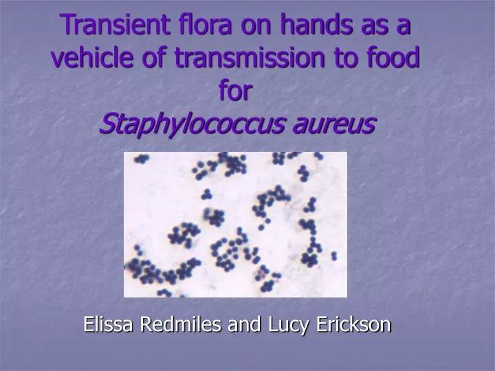
Transient flora on hands as a vehicle of transmission to food for Staphylococcus aureus. Elissa Redmiles and Lucy Erickson. Background. History of food bourn illness Hypothesis. History of food borne illness. Since the beginning of human history, food borne illness has been an issue

E N D
Transient flora on hands as a vehicle of transmission to food for Staphylococcus aureus Elissa Redmiles and Lucy Erickson
Background • History of food bourn illness • Hypothesis
History of food borne illness • Since the beginning of human history, food borne illness has been an issue • Various food preservation methods have been employed over the years to slow down food spoilage by microbes and natural aging processes, some common prevention methods include: • drying • freezing • freeze-drying • salting • curing • canning • pickling • irradiation • treating with sugar • treating with inert gases (such as carbon dioxide) • S
History of food borne illness (cont.) • Transient flora found on human skin and hands plays an important role in food contamination • When those involved in food preparation fail to observe hygienic methods
Hypothesis • It is hypothesized that Staphylococcus aureus, a microbe occurring naturally on the hands of some people, would be found at various locations throughout the Microbiology building • It was also hypothesized that many different types of bacteria would be isolated from the various locations tested
Hypothesis (cont.) • The isolation of bacteria from the inside handle of the bathroom door particularly holds implications for food hygiene, as ideally it should only be touched by pristinely washed hands.
Protocol Design and Detailed Methods • Overall approach and rationale for approach • Detailed methods and rationale behind methods • Expected results
Overall approach and rationale for approach • There were 2 objectives for this experiment: • To determine the number of different types of bacteria that live on each of 7 objects tested • To identify on which objects Staphylococcus aureus is present
Overall approach and rationale for approach (cont.) • To achieve these objectives a growth medium was required • Both agar and broth were considered • Agar was determined to be a more suitable growth medium since: • It allows direct observation of different types of bacterial colonies • It prohibits the number of bacteria on an object from being determined • Broth was determined to be a less suitable growth medium because: • It allows the number of bacteria on an object to be determined • It does not allow direct observation of different types of bacterial colonies
Overall approach and rationale for approach (cont.) • To achieve the first objective a sampling technique was required • Both saline wetted and dry sterile cotton swabs were considered: • Wet cotton swabs were determined to be most effective for obtaining the largest sample of microorganisms • Microorganisms adhere more readily to wet surfaces • A swab and streak method was used to inoculate the agar plates
Overall approach and rationale for approach (cont.) • To achieve the second objective a method of differentiating Staphylococcus aureus was needed: • Differential mediums and assays were used: • The gram stain was used to differentiate gram negative organisms from gram positive organisms • The catalase assay was used to differentiate Staphylococci (positive result) from Streptococci (negative result) • Blood Agar was used to differentiate S. aureus (positive result) from S. epidermis (negative result) • Mannitol Salt Agar was used to differentiate S. aureus (positive result) from S. epidermis (negative result) • The CoAgulase assay was used to differentiate S. aureus (positive result) from S. epidermis (negative result)
Detailed Methods • Day 1 • Made Mannitol Salt Agar • Autoclaved and cooled medium • Day 2 • Poured Mannitol Salt Agar plates • Day 3 • Obtained Trypticase Soy Agar (TSA) plates • Obtained a package of sterile cotton swabs, and a tube of diluted saline • Swabbed 7 objects: • Bathroom sink faucet (1st floor) • Bathroom (interior) doorknob (1st floor) • Micro bench sink faucet (Room x) • Front door handle—Micro building • Handicapped door button—Micro building • Computer mouse in lab • Stair rail (1st floor)
Detailed Methods (cont.) • Day 3 (cont.) • Swabbing method used: • Dipped tip of sterile swab into saline • Swabbed 1 of 7 objects • Struck inoculated swab on sterile agar plate with zigzag motion • A new swab was used for each plate • 4 plates (2 TSA and 2 Mannitol Salt Agar) were used for each object • Discarded the swab • Incubated all plates at 37°C for 48 hours
Detailed Methods (cont.) • Day 4 • Observed growth of different colony types on TSA plates • Observed growth of colonies on Mannitol Salt Agar plates • Observed fermentation • Photographed certain colonized plates • Counted and recorded different types of colonies • Selected different types of colonies from each Mannitol Salt Agar plate • Struck the selected colonies on (new) Mannitol Salt Agar plates, on (new) TSA plates, and on (new) Blood Agar plates • Incubated all plates at 37°C for 24 hours
Detailed Methods (cont.) • Day 5 • Observed Mannitol Salt Agar plates • Observed fermentation • Observed TSA plates • Observed Blood Agar plates • Observed hemolysis • Performed catalase assay on pure colonies isolated from Mannitol Salt Agar plates • Performed gram stain on pure colonies isolated from Mannitol Salt Agar plates • Performed CoAgulase assay on certain pure colonies isolated from Mannitol Salt Agar plates • Bactistaph latex 150 test • Photographed relevant results • Day 6 • Observed results from CoAgulase assay
Detailed Rationale Behind Methods • Day 3 • Saline was used: • To maintain highest degree of sterility • Since bacteria adhere more readily to wet surfaces than to dry surfaces • In a diluted form so growth of non-salt tolerant organisms was uninhibited • A new swab was used for each plate to reduce the risk of contamination by organisms not found on objects of interest • TSA plates were used: • As a control • To determine the different types of organisms on objects of interest
Detailed Rationale (cont.) • Day 3 (cont.) • Mannitol Salt Agar plates were used: • To isolate Staphylococcus aureus from objects of interest • Staphylococcus aureus is a salt tolerant organism that grows on Mannitol Salt Agar while other non-salt tolerant organisms will not grow • All plates were incubated at 37°C for 48 hours: • 37°C is in the optimal growth temperature range for organisms that could be found on objects of interest • 48 hours is in the optimal growth time range for organisms that could be found on objects of interest • 4 plates (2 TSA and 2 Mannitol Salt Agar) were used: • To safeguard from error • If an error occurred with one “set” (1 TSA and 1 Mannitol Salt Agar plate) results could be obtained from another “set” (theoretically void of error)
Detailed Rationale (cont.) • Day 4 • Different colonies from each TSA plate were struck on (new) TSA plates to confirm that colonies selected were pure and different from other colonies on the plate • Struck colonies from Mannitol Salt Agar on (new) Mannitol Salt Agar plates to isolate pure colonies for further tests and to confirm that colonies isolated were Staphylococcus aureus • Staphylococcus aureus ferments Mannitol producing a yellow zone surrounding colony
Detailed Rationale (cont.) • Day 4 (cont.) • Struck colonies from Mannitol Salt Agar on (new) TSA plates as a control • Isolated colonies were struck on Blood Agar to confirm that isolated colonies were Staphylococcus aureus • Staphylococcus aureus has -hemolysis on Blood Agar
Detailed Rationale (cont.) • Day 5 • Catalase test was performed to: • Confirm that cultures from Mannitol Salt Agar plates were Staphylococci (positive) not Streptococci (negative) • Indicating that cultures were Staphylococcus aureus • Gram stain was performed to: • Confirm that cultures from Mannitol Salt Agar plates were gram positive not gram negative • Indicating that cultures were Staphylococcus aureus • Confirm that bacteria colonized on Mannitol Salt Agar plates formed clusters not chains • Indicating that cultures were Staphylococcus aureus
Detailed Rationale (cont.) • Day 5 (cont.) • Bacti Staph Latex 150 test was performed to: • Confirm that isolated colonies tested positive not negative for agglutination • Indicating that the cultures were Staphylococcus aureus • CoAgulase assay was performed to: • Confirm that isolated colonies were CoAgulase positive not CoAgulase negative • Indicating that the cultures were Staphylococcus aureus
Expected Results • It was expected that: • Numerous types of microorganisms would live on the tested objects • Staphylococcus aureus is found on some human hands and would therefore be present on some of the tested objects
Results • Findings • Summary of Findings • Relationship of findings to expectations
Findings • TSA plate 1 was inoculated with microorganisms from the bathroom sink faucet: • 7 small white colonies grew • 8 large white colonies grew • TSA plate 2 was inoculated with microorganisms from the bathroom sink faucet: • 16 small white colonies grew • 31 large white colonies grew • Mannitol Salt Agar plate 1 was inoculated with microorganisms from the bathroom sink faucet: • 2 small white colonies grew • Mannitol Salt Agar plate 2 was inoculated with microorganisms from the bathroom sink faucet: • No growth
Findings (cont.) • TSA plate 1 was inoculated with microorganisms from the computer mouse: • No growth • TSA plate 2 was inoculated with microorganisms from the computer mouse: • 1 small white colonies grew • 1 medium white colonies grew • Mannitol Salt Agar plate 1 was inoculated with microorganisms from the computer mouse: • 2 small white colonies grew • 3 large peaked colonies grew • Mannitol Salt Agar plate 2 was inoculated with microorganisms from the computer mouse: • No growth
Findings (cont.) • TSA plate 1 was inoculated with microorganisms from the stair rail: • No growth • TSA plate 2 was inoculated with microorganisms from the stair rail: • No growth • Mannitol Salt Agar plate 1 was inoculated with microorganisms from the stair rail: • 1 small white colony grew • 1 large colony with yellow zone grew • Mannitol Salt Agar plate 2 was inoculated with microorganisms from the stair rail: • No growth
Findings (cont.) • TSA plate 1 was inoculated with microorganisms from the handicapped button: • 1 small yellow colony grew • TSA plate 2 was inoculated with microorganisms from the handicapped button: • 1 small white colony grew • Mannitol Salt Agar plate 1 was inoculated with microorganisms from the handicapped button: • No growth • Mannitol Salt Agar plate 2 was inoculated with microorganisms from the handicapped button: • No growth
Findings (cont.) • TSA plate 1 was inoculated with microorganisms from front door knob: • 33 small yellow colonies grew • 1 medium white colony grew • TSA plate 2 was inoculated with microorganisms from the front door knob: • 1 small white colony grew • Mannitol Salt Agar plate 1 was inoculated with microorganisms from the front door knob: • No growth • Mannitol Salt Agar plate 2 was inoculated with microorganisms from the front door knob: • No growth
Findings (cont.) • TSA plate 1 was inoculated with microorganisms from bathroom door knob: • 3 small yellow colonies grew • 1 small white colony grew • 1 medium white colony grew • TSA plate 2 was inoculated with microorganisms from the bathroom door knob: • 7 small white colonies grew • 9 large white colonies grew • Mannitol Salt Agar plate 1 was inoculated with microorganisms from the bathroom door knob: • 2 medium colonies with yellow zones grew • 2 large peaked colonies grew • Mannitol Salt Agar plate 2 was inoculated with microorganisms from the bathroom door knob: • No growth
Findings (cont.) • TSA plate 1 was inoculated with microorganisms from lab sink: • No growth • TSA plate 2 was inoculated with microorganisms from the bathroom door knob: • No growth • Mannitol Salt Agar plate 1 was inoculated with microorganisms from the bathroom door knob: • 1 small white colony grew • Mannitol Salt Agar plate 2 was inoculated with microorganisms from the bathroom door knob: • No growth
Findings (cont.) • A Mannitol Salt Agar plate was inoculated with microorganisms from the bathroom door handle: • Growth and fermentation on Mannitol plate • Gram positive rods • Snapping replication was observed • Catalase positive • A Mannitol Salt Agar plate was inoculated with microorganisms from the bathroom sink: • Growth and fermentation on Mannitol plate • Gram positive cocci organized in grape like clusters • Catalase positive
Findings (cont.) • A Mannitol Salt Agar plate was inoculated with microorganisms from the stair rail: • Growth and fermentation on Mannitol plate • Gram positive rods • Snapping replication was observed • Catalase negative • A Mannitol Salt Agar plate was inoculated with microorganisms from the computer mouse: • Growth and fermentation on Mannitol plate • Gram positive rods • Snapping replication was observed • Catalase positive
Findings (cont.) • A Blood Agar plate was inoculated with microorganisms from the bathroom door handle: • Growth on Blood Agar • β-hemolysis • A Blood Agar plate was inoculated with microorganisms from the bathroom sink: • Growth on Blood Agar • β -hemolysis • A Blood Agar plate was inoculated with microorganisms from the stair rail: • Growth on Blood Agar • β-hemolysis • A Blood Agar plate was inoculated with microorganisms from the computer mouse: • Growth on Blood Agar • α-hemolysis
Summary of Findings • Microorganisms isolated from the objects exhibited a variety of phenotypes when grown on TSA plates: • Small white bacterial colonies • Small yellow bacterial colonies • Medium white bacterial colonies • Large white bacterial colonies • The phenotypes above indicate that a very diverse bacterial population is present on the tested objects • This data supports the first hypothesis that numerous types of microorganisms would live on tested objects
Summary of Findings (cont.) • Microorganisms isolated from the: • Stair rail • Computer mouse • Bathroom door handle • Are suspected to be Cornyebacteria since: • They undergo snapping replication • Form “Chinese letters” • Are gram positive rods • This was not hypothesized
Summary of Findings (cont.) • Microorganisms isolated from the bathroom sink are suspected to be Staphylococcus aureus • The samples are strongly suspected to be Staphylococcus aureus since: • They are gram positive cocci seen in grape-like clusters • Are catalase and CoAgulase positive • Have β-hemolysis • Ferment Mannitol • These phenotypes are identical to those exhibited by Staphylococcus aureus • This supports the second hypothesis: Staphylococcus aureus is found on human hands and would therefore be present on some of the tested objects
Discussion • Description of Staphylococcus aureus • Why Staphylococcus aureus was expected to be found • Incidence of disease • Virulence of Staphylococcus aureus • Comments on protocol • Comments on results • Comments on overall significance of project
Description of Staphylococcus aureus • Phenotype • Gram positive cocci • Found in pairs, chains, or grapelike-clusters • Produces heat stable enterotoxin • Salt tolerant • Ferments Mannitol • Catalase positive • CoAgulase Positive
Description of Staphylococcus aureus (cont.) • Habitat • Air • Dust • Sewage • Environmental surfaces • 50% of humans (higher in health professionals) • Foods • Poultry and egg products • Egg, tuna, potato, and chicken salads • Milk and dairy products
Why Staphylococcus aureus was expected to be found • Expected to findStaphylococcus aureus various surfaces throughout the microbiology building where the transient flora from many peoples’ hands has been transferred. One of the places swabbed was the inside bathroom door handle, where hopefully only people with freshly washed hands are touching. Unfortunately, this is not always the case, and a diverse population of bacteria were isolated from the door handle. Someone who fails to wash his or her hands upon exiting the restroom is unlikely to remember to wash them before eating or preparing food for another person. Thus, if S. aureus were found on the locations tested it could hold strong ramifications for general human cleanliness, and subsequently, health.
Why finding Staphylococcus aureus was probable • Staphylococcus aureus is a component of the normal flora for 50% of all people • Carriers of Staphylococcus aureus transfer the bacteria to every touched object • Regardless of hand washing • In proper growth conditions the bacteria will propagate on objects such as those tested • As Staphylococcus aureus contaminated objects are used, bacteria are transferred to a users transient flora • Staphylococcus aureus was isolated on tested objects • Thus, hand washing does not preclude the transfer of Staphylococcus aureus to food
Incidence of disease • The incidence of illness is difficult to pinpoint due to a number of reasons • Poor response from victims during interviews • Misdiagnosis (symptoms are very similar to those of Bacillus cereus toxin) • Inadequate collection of lab samples • Improper lab examination
Virulence of Staphylococcus aureus • Causes staphyloenterotoxicosis (food poisoning) • Operates by toxin, not colonization • ID50 – 1.0 microgram • Onset of illness is rapid and varies based on: • Amount of food ingested • Amount of toxin present in food • Individual susceptibility • General health of victim
Virulence of Staphylococcus aureus (cont.) • Symptoms: • Nausea • Vomiting • Abdominal cramping • Headache • Changes in pressure and blood rates • Average recovery time for mild case: • 2-3 days
Virulence of Staphylococcus aureus (cont.) • Treatment • Rest • Fluids • Medicines to calm stomach • Hospitalization/Intravenous therapy for more severe cases (usually elderly or infants)
Virulence of Staphylococcus aureus (cont.) • Prevention • Wash hands and under nails vigorously and often • Do not prepare food with an eye or nose infection • Do not serve food to others with open wounds • Keep food preparation areas sanitary • Store cooked food in wide, shallow containers and refrigerate • Do not leave food out for long periods of time • Keep hot foods hot (above 140 degrees F) • Keep cold foods cold (40 degrees F or lower)
Comments on Protocol • Method of multiple plate inoculation was successful • By using 4 plates for each object tested it allowed for confirmation of results • Saline wetted swab and streak inoculation method was successful: • Isolated colony growth occurred • Testing and differential media: • Catalase • CoAgulase • Mannitol • Blood
Comments on Results • Comments on Results • Found numerous colonies resembling Corynebacteria, a Gram-Positive rod generally found in soil • Methylene blue staining did not reveal metachromatic granules, however not all species exhibit this trait • This finding is consistent with concept of bacteria found on unwashed hands • Unable to identify specific strain of Corynebacteria isolated, since many of the organisms can not be typed easily. Although there have been significant advances in PCR technology, this technology was not available. • Isolate from bathroom sink faucet is indicated by tests to be Staphylococcus aureus
Comments on overall significance of project • This project aims to raise public awareness that pathogenic microorganisms are present on numerous objects touched prior to and after hand washing
References • Boyce, J., Pittet, D. (2002). “Guideline for Hand Hygiene in Health-Care Settings” 1 April 2006. Morbidity and Mortality Weekly Report, 51-RR16. 1-44 <http://www.cdc.gov/mmwr/preview/mmwrhtml/rr5116a1.htm> • CDC. Outbreak of community-associated methicillin-resistant Staphylococcus aureus skin infections---Los Angeles County, California, 2002--2003. Morbidity and Mortality Weekly Report 2006;52:88. <http://www.cdc.gov/mmwr/preview/mmwrhtml/mm5512a1.htm> • U.S. Food and Drug Administration. “Staphylococcus aureus.” 1992. Food borne Pathogenic Microorganisms and Natural Toxins Handbook. <http://www.cfsan.fda.gov/~mow/chap3.html>