Download
1 / 42
500 likes | 912 Views
INTERSTITIAL AND INFILTRATIVE PULMONARY DISEASES ( DIFFUSE PARENCHYMAL LUNG DISEASE ) (Restrictive pulmonary Diseases). INTRODUCTION. Restrictive lung diseases: Intrinsic lung diseases : alteration in lung parenchyma

E N D
INTERSTITIAL AND INFILTRATIVE PULMONARY DISEASES ( DIFFUSE PARENCHYMAL LUNG DISEASE ) (Restrictive pulmonary Diseases)
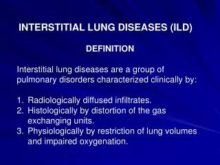
INTRODUCTION • Restrictive lung diseases: • Intrinsic lung diseases: alteration in lung parenchyma • Extrinsic lung diseases: diseases of pleura, chest wall, or neuromuscular apparatus.
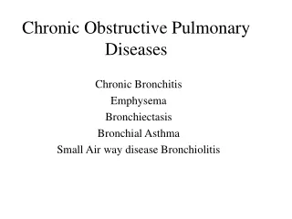
Restrictive Pulmonary Disease Indicate limitation to full expantion of the lungs because of diseases in the lung paranchyma , chest wall or diaphragm. ( Lung volumes are decrease but flow rates are normal ). As Interstitial lung disease( cryptogenic fibsosing alveolitis , sarcoidosis, asbestosis, silicosis , coal worker pneumoconiosis…) TLC ( total lung capacity ) decrease RV ( residual volume ) decrease FEV1 / VC = or more than 70%
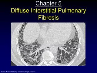
Overview of interstitial lung diseases The interstitial lung diseases (diffuse parenchymal lung diseases DPLDs) are a heterogeneous group of conditions caused by diffuse thickening of the alveolar wallswith inflammatory cells and exudates (e.g. the acute respiratory distress syndrome-ARDS), granulomas (e.g. sarcoidosis), alveolar haemorrhage (e.g. Goodpasture's syndrome), and/or fibrosis (e.g. fibrosingalveolitis). Lungs start to scar and the interstitium(tissue between the air sacs) and lungs become stiff .
Interstitial lung diseases • The interstitial lung diseases (diffuse parenchymal lung diseases DPLDs) are a heterogeneous group of conditions caused by several different pathological processes , having similar symptomes , signs , radiological changes & pulmonary function tests. • The pathological changes are : 1- Thickening of alveolar wall by oedema, cellular exudates or fibrosis. 2- Increased stiffness of the lungs( reduced compliance).causing exertional dyspnoea. 3- Maldistribution of ventilation & perfusion, & gas transfer defect.causing hypoxemia particularly on exertion.
causes of interstitial lung diseases 1- Collagen vascular diseases • Scleroderma • Polymyositis/dermatomyositis • Systemic lupus erythematosus • Rheumatoid arthritis • Ankylosing spondylitis.
2- Drugs induced • Nitrofurantoin • Amiodarone • Gold • Bleomycin • Cyclophosphamide • Methotrexate • Radiation.
3- Primary or unclassified diseases • Sarcoidosis • Pulmonary histiocytosis X • Lymphangioleiomyomatosis (LAM) • Pulmonary vasculitis • Alveolar proteinosis • Eosinophilic pneumonia • Bronchiolitis obliterans organizing pneumonia (BOOP)
4- Idiopathic fibrotic disorders • Acute interstitial pneumonia • Idiopathic pulmonary fibrosis (usual intersttial pneumonitis) • Lymphocytic interstitial pneumonitis • Desquamative interstitial pneumonitis • Non-specific interstitial pneumonitis
5- Inhaled inorganic dust • Silica: silicosis • Asbestos: asbestosis • Beryllium: berylliosis • Cobalt: hard metal fibrosis • Coal workers pneumoconiosis 6- Inhaled organic dust • Hypersensitivity pneumonitis: farmer’s lung Bird fanciers lung….
7- Others -As part of systemic inflammatory diseases e.g.ARDS. -Chronic pulmonary oedema e.g. secondary to mitral srenosis.
Diagnosis of INTERSTITIAL LUNG DISEASES • Diagnosis often presents a considerable clinical challenge , necessitating meticulous attention to the historyand physical signs and a cooperative approach from teams of clinicians, radiologists and pathologists.( investigations )
History • The duration of disease may sometimes be difficult to ascertain. • Gradually progressive shortness of breath on exertion may be the only symptom, and hence the patient may not present clinically until there is extensive lung pathology. • History-taking should include a thorough and comprehensive search for exposure to organic and inorganic dusts. • A 'lifetime' occupational history is essential and should include hobbies that may involve similar exposures.
Contact with birds at home or in the working environment is the cause of the most common form of hypersensitivity pneumonitis (HP). • The smoking status should be recorded and a drug history should be obtained. • A history of rashes, joint pains or renal disease may suggest an underlying connective tissue disorder or vasculitis . The presence of any comorbid disease should be ascertained such as collagen vascular disease, immunodeficiency, HIV or malignancy. • In exceptional cases there is a family history of DPLD.
Clinical features of interstitial lung diseases • Gradually progressive SOB on exertion & exercise limitation. • Dry non productive cough. • Digital clubbing (as in fibrosing alveolitis & asbestosis). • Restriction of chest expantion. • Auscultation: end inspiratory crepitations particularly over the lower zones posteriorly & laterally. In advanced cases:. tachypnoea , cyanosis , signs of pulmanary hypertention & Rt. Heart failure Extrapulmonary signs, including lymphadenopathy or uveitis, may be present in sarcoidosis arthropathies or rashes may occur when a DPLD is a manifestation of a connective tissue disorder .
Signs and Symptoms • Breathlessness • Dry cough • Fingertips enlarge and nails curve over tops of fingertips
Investigations in interstitial lung diseases Laboratory investigations . Full blood count-lymphopenia in sarcoid; eosinophilia in pulmonary eosinophilias and drug reactions; neutrophilia in hypersensitivity pneumonitis • Ca2+-may be elevated in sarcoid • Lactate dehydrogenase (LDH)-may provide non-specific indicator of disease activity . • Serum ACE-non-specific indicator of disease activity in sarcoid • ESR and CRP may be non-specifically raised • Autoimmune screen and rheumatoid factor may suggest collagen vascular disease Radiology . Chest X-ray • High-resolution CT scan • Gallium scanning
Chest X-ray typically shows a fine reticular, reticulonodular or even nodular pattern of infiltration at the bases and periphery with cystic areas and honeycombing in advanced disease. However, plain radiography is insensitive and may not appear abnormal until disease is advanced. HRCT is more sensitive and specific and has become extremely valuable in detecting early interstitial lung disease, assessing the extent and type of involvement and guiding further investigations and management
Pulmonary function Spirometry, lung volumes, gas transfer. Bronchoscopy Bronchoalveolar lavage, differential cell counts Bronchial biopsy may be useful in sarcoid . Lung biopsy (in selected cases) Transbronchial biopsy useful in sarcoid and differential of malignancy or infection Video-assisted thoracoscopy (VATS) Others Liver biopsy Urinary calcium excretion may be useful in sarcoid
Pneumoconiosis Lung disease caused by mineral dust exposure: • Asbestosis • Coal workers lung • Silicosis
IDIOPATHIC INTERSTITIAL PNEUMONIAS • The idiopathic interstitial pneumonias (IIPs) are characterised by varying patterns of inflammation and fibrosis in the lung parenchyma. Idiopathic interstitial pneumonias include the following diseases: -Idiopathic pulmonary fibrosis (IPF). (The most important disease ) - desquamative interstitial pneumonia (DIP), - acute interstitial pneumonia (AIP), - non-specific interstitial pneumonia (NSIP), -respiratory bronchiolitis-interstitial lung disease (RB-ILD) -cryptogenic organising pneumonia (COP) -lymphocytic interstitial pneumonia (LIP)
IDIOPATHIC PULMONARY FIBROSIS (IPF) This term has replaced cryptogenic fibrosing alveolitis and refers to a specific form of DPLD characterised by pathological (or radiological) evidence of usual interstitial pneumonia (UIP). (IPF,CFA, UIP)
Aetiology of IPF • The aetiology remains unknown • speculation has included exposure to infectious agents such as Epstein-Barr virus, occupational dusts such as metal or wood dusts, prior use of antidepressants, and a possible role for chronic gastro-oesophageal reflux. • Familial cases are rare but genetic factors that control the inflammatory and fibrotic response are likely to be important. • The disease displays a strong association with cigarette smoking
IPF(CFA )has an incidence of 6 – 10 per 100 000 per year & is about twice as common among cigarette smokers as in non-smokers. • Men are more commonly affected than women. • Macroscopically , the lungs show subpleural fibrosis & a honeycomb appearance , predominantly in the lower lobes & basolateral pleural regions. • Microscopically , there are fibroproliferative lesions representing the site of healing alveolar injury. • there is a variable mononuclear cell infiltration of the alveolar walls , fibrosis & smooth muscle proliferation.
Clinical features of IPF • CFA is a disease of the elderly , with a mean age at presentation of 69 years. • Progressive exertional breathlessness is usually the presenting symptom, often accompanied by dry cough. • Digital clubbing is observed in 60 % of cases. • Chest expantionis poor. • On chest auscultation : bilateral end – inspiratory crepitations ,particularly over the lower zones posteriorly.
Investigations of IPF 1- Blood tests • are of little value. • Rheumatoid factor and antinuclear factor can be detected in 30-50% of patients. • The erythrocyte sedimentation rate (ESR) and lactate dehydrogenase (LDH) are elevated in most cases.
2- Radiological investigations: CXR • diffuse pulmonary opacities more obvious in the lower zones & peripherally.( lower zone bi-basal reticular and reticulonodular opacities). • The hemidiaphragms are high & the lungs appear small. • In advanced cases CXR may shows a 'honeycomb' appearance ( in which diffuse pulmonary shadowing is interspersed with small cystic translucencies) but it is non-specific . HRCT • may be diagnostic, demonstrating a patchy, predominantly peripheral, subpleural and basal reticular pattern with subpleural cysts (honeycombing) and/or traction bronchiectasis and is particularly useful in early disease when chest X-ray changes may be indistinct.
Honeycomb lung • Subpleural “enlarged” spaces with fibrous walls
3- Pulmonary function tests: show a restrictive defect with reduced lung volumes and gas transfer. TLC ( total lung capacity ) decrease RV ( residual volume ) decrease FEV1 / VC = or more than 70% gas transfer factor decrease. (However, lung volumes may be preserved in patients with concomitant emphysema).
4- Arterial blood gases: - In early stages : arterial hypoxaemia on exertion. - In later stages : arterial hypoxaemia & hypocapnia at rest.
5- Lung biopsy: • A firm diagnosis of (CFA IPF, UIP) can usually be achieved on the bases of the history , clinical finding & HRCT , But if doubt exist an open lung biopsy is indicated. (Patients with typical clinical features and HRCT appearances consistent with UIP do not require lung biopsy, particularly if other known causes of interstitial lung disease have been excluded). 6- Bronchoalveolarlavage: may shows an increased number of neutrophils. • Transbronchial biopsy & Bronchoalveolarlavage may be used to exclude alternative diagnoses
1- Immunosupressive therapy: Corticosteroid :the response to steroid is usually poor, but a proportion of patients do respond in terms of symptoms(50%) & lung function(25%). Indications for using corticosteroid: a- Highly symptomatic patient. b- Rapidly progressive disease. c- Patient who have a predominantelyground glass appearance on HRCT. d- Fall of more than 15% in FVC (Forced Vital Capacity ) or gas transfer over 3 – 6 month period. The initial treatment is combined therapy with Prednisolone(0.5 mg/kg) combined with azathioprine(2-3 mg/kg). Assessment of response to this treatment is by repeated measurment of lung volumes , gas transfer facror & CXR. Immunosuppressive therapy should be withdrown over a few weeks if there is no response. If there is objective evidence of improvement , the prednisolone dose can be reduced gradually to a maintenance dose of 10 – 12.5 mg / day.
2-Lung transplantation : should be considered in young patients with advanced disease. 3- Others: Cyclophosphamideoccasionally used. Antifibrotics such as colchicinemay be used. Oxygen, rehabilitation, psychosocial aspects are helpful
Prognosis of IPF • CFA has a high mortality rate . The 5 year survival rate is less than 30 % • The median survival time of patients with CFA is about 3.5 years • Most deaths occur in patients over the age of 55 years with male predominating. • In the majority of patients the disease is progressive , even in those who have responded to treatment. • High numbers of fibroblastic foci on biopsy have been associated with a poor outcome.