Download
1 / 49
520 likes | 1.01k Views
CNS TUBERCULOSIS IMAGING AND SURGERY. Tuberculosis . As old as recorded history Symptoms described in the Rig Veda (1500 BC) Unequivocal lesions in Egyptian mummies Odier, Ford described meningeal TB 1790 Surgical excision Wernicke and Hahn 1882, . Tuberculosis .

E N D
Tuberculosis • As old as recorded history • Symptoms described in the Rig Veda (1500 BC) • Unequivocal lesions in Egyptian mummies • Odier, Ford described meningeal TB 1790 • Surgical excision Wernicke and Hahn 1882, CNS tuberculosis imaging and surgery
Tuberculosis • CNS tuberculosis complicates 10% of all TB • Never the first manifestation • Occurs within 6-12 months • Circle of Willis more frequently involved than the basilar system CNS tuberculosis imaging and surgery
Mycobacterium tuberculosis • Acid fast bacillus • Does not stain on gram stain • Obligate aerobes • Difficult to grow • High lipid in cell wall • Hominis/ Bovine/ Avium CNS tuberculosis imaging and surgery
Pathogenesis • May develop during initial infection/ reactivation • Haematogenous dissemination • Commonest • Focus in brain (Rich focus) • Rupture of focus into subarachnoid/ ventricular space • Contiguous spread CNS tuberculosis imaging and surgery
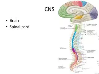
CNS tuberculosis • Intracranial • Parenchymal • Meningeal • Osseous • Spinal • Parenchymal • Meningeal • Arachnoiditis • Osseous CNS tuberculosis imaging and surgery
Epidemiology • Incidence varies blacks > whites • Predominantly in the young (50% <10) • Abscess in 4-8% (20% with HIV) CNS tuberculosis imaging and surgery
Pathology • Immature lesions – multiple tubercles in oedematous brain • Mature: avascular mass, nodular extensions, yellowish gritty casseous areas • 60% attached to dura CNS tuberculosis imaging and surgery
Pathology (parrenchymal) • Can be present anywhere • Cerebellum in children • Cerebral hemisphere and basal ganglia commoner in adults CNS tuberculosis imaging and surgery
Pathology (tuberculoma) • Tuberculoma ( classical lesion) • Tuberculoma en plaque • Tuberculous abscess • Cystic tuberculoma • Multiple grape like tuberculoma • Microtuberculoma • Calcified tuberculoma • Tuberculous encephalopathy CNS tuberculosis imaging and surgery
Pathology (tuberculoma) • Dastur described six main types • Parenchymal changes. • (1) Ventriculitis • (2) Border-zone encephalitis • (3) Infarction • (4) Internal hydrocephalus • (5) Diffuse oedema • (6) Tuberculoma CNS tuberculosis imaging and surgery
Pathology (meningeal) • Classically Commonest in 6m – 3 years • Now adults 50% • Thick exudate encasing nerves, vessels • HCP, tuberculoma, arachnoiditis • Diffuse perivasculitis • Infarcts • Pachymeningitis CNS tuberculosis imaging and surgery
Diagnosis • Montoux test • Hb/ ESR • CXR • ELISA • CSF • PCR • Imaging • Biopsy CNS tuberculosis imaging and surgery
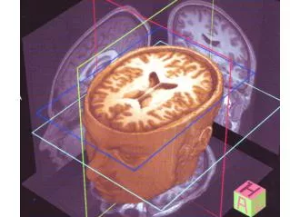
Imaging • X ray • Angiography of historical significance • CT • MRI CNS tuberculosis imaging and surgery
Imaging • Tuberculoma • Typically cortical and subcortical • Multiple in 10-35% • Milliary rare ( children) • Meningitis (commonest form of CNS TB) • Isolated meningitis is rare (5% in children) • Basal cisterns CNS tuberculosis imaging and surgery
Imaging (CT tuberculoma) • Cerebritis: hypodense areas • Perilesionaloedema out of proportion • Early tuberculoma: iso to slightly hyper dense , ring enhancement • Evolved : well delineated ring enhancing mass, target sign (central enhancement or calcification) • Healed: often calcify • Manifestations • Small disc/ rings • Large rings with central lucency • Large nodular mass with irregular outline • Multiple lesions in 15-20% CNS tuberculosis imaging and surgery
Caseating tuberculosis granuloma involving the right frontal lobe. CECT shows a rim-enhancing lesion in the right frontal lobe consistent with a caseating tuberculosis granuloma CNS tuberculosis imaging and surgery
Imaging (MRI tuberculoma) • T1 : isointence • T2: central hyper with hypo ring • Marked thin rim enhancement • Hypo on T2: fibrosis, gliosis, macrophage infiltration CNS tuberculosis imaging and surgery
Parrenchymal tuberculosis. contrast-enhanced T1-weighted MR image demonstrates multiple enhancing caseating and non-caseating tuberculomas, predominantly within the left frontal and parietal lobes CNS tuberculosis imaging and surgery
Milliary CNS tuberculosis. Axial contrast-enhanced T1-weighted MR image shows multiple small high-signal-intensity foci within both cerebral hemispheres CNS tuberculosis imaging and surgery
A. Bernaerts, F. M. VanhoenackerTuberculosis of the central nervous system: overview of neuroradiological findings. Eur Radiol (2003) 13:1876–1890 CNS tuberculosis imaging and surgery
Imaging (meningitis) • Active • Sequelae • Hydrocephalus • Ischemia and infarction • Medial lenticulostriate 75% • Thalamoperforating • Cortex 25% • Bilateral 70% • Atrophy • Calcification CNS tuberculosis imaging and surgery
Imaging (CT meningitis) • NCCT: • scans may be normal • Obliteration of basal cisterns by hypo/ iso dense exudate • en plaque dural thickening • Popcorn calcification • Hydrocephalus • Sequelae of chronic meningitis • Infarcts • CECT: • Abnormal meningeal enhancement (may persist) • Leptomeningeal enhancement sylvian fissures, tentorium • Granulomas in the basal meninges • Ependymitis CNS tuberculosis imaging and surgery
Imaging (MRI meningitis) • Unenhanced scan: does not show active meningitis • Spine • CSF loculations • Obliteration of arachnoid space • Loss of cord outline in cervicodorsal cord • Thickening and clumping of roots in the lumbar cord • Contrast T1 : basal meningeal enhancement • spine • Linear enhancement of cord/ roots CNS tuberculosis imaging and surgery
Tuberculous meningitis. Axial contrast-enhanced T1-weighted magnetic resonance (MR) image shows florid meningeal enhancement. CNS tuberculosis imaging and surgery
Tubercular meningitis. Axial FLAIR-MR] showing marked hyperintensity of the basal cisterns and prominent temporal horns in a patient with mild communicating hydrocephalus CNS tuberculosis imaging and surgery
Tubercular spondylitis with epidural and retroabscess CNS tuberculosis imaging and surgery
Enhanced T1-weighted magnetic resonance imaging with fat suppression show intense enhancement of the subarachnoid space indicating arachnoiditis CNS tuberculosis imaging and surgery
Tuberculous pachymeningitis • Rare • Common sites of involvement are cavernous sinus, floor of middle cranial fossa and tentorium. • Radiographic features • CT hyperattenuating solid plaque like densities (calcification may be seen) • MRI • T1 : hypo intense thickened duramater. • T2 : hypo intense thickened meninges. • T1 C+ (GAD) : intense homogenous enhancement of thickened meninges. CNS tuberculosis imaging and surgery
Management • Medical therapy • Surgery • indications • Vision or life threatened by mass effect • Failure of response to medical therapy • Paradoxical increase in lesion size with therapy • Diagnosis in doubt CNS tuberculosis imaging and surgery
Medical therapy CNS tuberculosis imaging and surgery
WHO recommendations • PULMONARY AND EXTRA PULMONARY DISEASE SHOULD BE TREATED WITH SAME REGIMENS. NOTE THAT SOME EXPERTS RECOMMEND 9-12 MONTHS OF TREATMENT OD TB, MENINGITIS (2,3)GIVEN THE SERIOUS RISK OF DISABILITY AND MORTALITY, AND 9 MONTHS OF TREATMENT FOR TB OF BONES OR JOINTS, BECAUSE OF DIFFICULITIES OF ASSESING TREATMENT RESPONSE (3).UNLESS DRUG RESISTANCE IS SUSPECTED, ADJUVENT CORTICOSTERIODS TREATMENT IN RECOMMENDED FOR TB MENINGITISAND PERICARDITIS(1-4). IN TUBERCULOUS MENINGITIS, ETHAMBUTOL SHOULD BE REPLACED WITH STREPTOMYCIN. 2. National collabrating centre for chronic conditions. Tuberculosis : clinical diagnosis and management of tuberculosis, measures of its preventions and control. London royal college of physicians, NICE, 2006. 3. American thoracic society , CDC, infectious disease society of America. Treatment of tuberculosis morbidity and mortality weekly report: recommendations and reports,2003, 52(R-11):1-77. WHO Treatment of tuberculosis: guidelines – 4th ed CNS tuberculosis imaging and surgery
Duration of treatment 6 months van Loenhout-Rooyackers JH, Keyser A, Laheij RJ, Verbeek AL, van der Meer JW. Tuberculous meningitis: Is a 6-month treatment regimen sufficient? Int J Tuberc Lung Dis 2001;5:128-35. 12 months Thwaites GE, Hein TT. Tuberculous meningitis: Many questions, too few answers. Lancet Neurol 2005;4:160-70 18 months or Longer Santosh Isac Poonnoose, Vedantam Rajashekhar: Rate of Resolution of histologically verified intracranial tuderculomas. Neurosurgery 53:873-879, 2003 CNS tuberculosis imaging and surgery
Treatment Rate of radiological resolution of intracranial tuberculoma Series duration of ATT residual lesions % Wang 1996 (16) 6 20 Rajeshwari 1995 (6) 9 12 Awada 1998 (2) 12 0 Poonnoose 2003 (28) 18 69.2 Santosh Isac Poonnoose, Vedantam Rajashekhar: Rate of Resolution of histologically verified intracranial tuderculomas. Neurosurgery 53:873-879, 2003 CNS tuberculosis imaging and surgery
Medical management • 4 drugs x 3-4 months • 2 drugs x 14-16 months occasionally longer • Regression of size from 4-6 weeks • Most resolve in 12-14 months R Patir, R Bhatia, Tandon PN. Surgical management of tuberculous infections of the nervous system. Schmidek and Sweet operative neurosurgical techniques 5th edition; 1617-1631 • AED to continue • INH blocks phenytoin metabolism • Steroids in all irrespective of age and stage Prasad K, Singh MB. Corticosteroids for managing tuberculous meningitis. Cochrane Database Syst Rev 2008;1:CD002244. CNS tuberculosis imaging and surgery
Resistant tuberculosis • MDR : resistant to INH and Rifampicin • EDR/ XDR : MDR + resistance to Quinolones and injectable second line drugs CNS tuberculosis imaging and surgery
Surgery • Severe elevation of ICP • Threatening life or vision • Do not respond to drugs clinically/ radiologically • Diagnosis in doubt • Obstructive hydrocephalus R Patir, R Bhatia, Tandon PN. Surgical management of tuberculous infections of the nervous system. Schmidek and Sweet operative neurosurgical techniques 5th edition; 1617-1631 • Aim diagnosis/ relieve pressure CNS tuberculosis imaging and surgery
Surgical management • Biopsy of the mass lesion • Hydrocephalus • Communicating (commoner) • Non communicating CNS tuberculosis imaging and surgery
Surgery principles • Non eloquent areas total excision (small lesion) • Subtotal/ partial excision (large lesion/ eloquent cortex) • Conservative excision around vital structures • Evacuation of central liquifactive portion in deep seated lesions • Residual lesions may respond to medical therapy • R Patir, R Bhatia, Tandon PN. Surgical management of tuberculous infections of the nervous system. Schmidek and Sweet operative neurosurgical techniques 5th edition; 1617-1631 • Hydrocephalus CNS tuberculosis imaging and surgery
Hydrocephalus • Inevitable in those who survive 4-6 weeks • Mortality 20-100% • Grade at admission significant • Early shunt for grade I,II CNS tuberculosis imaging and surgery
ETV • 73.1% success rate for ETV in TBM with hydrocephalus • A chugh, M hussain et al. Surgical outcome of tuberculous meningitis hydrocephalus treated by endoscopic third ventriculostomy: prognostic factors and postoperative neuroimaging for functional assessment of ventriculostomy: J Neurosurg Pediatrics 3:000–000, 2009 • Endovascular revascularization for ischemia • STA MCA bypass • The left superficial temporal artery–MCA bypass was found to be capable of preventing new ischemic events in the 21-month follow-up period • Martin misch, Ultrich- wilhelm et al. Prevention of secondary ischemic events by superficial temporal artery–middle cerebral artery bypass surgery after tuberculosis-induced vasculopathy in a 5-year-old child:Neurosurg Pediatrics 6:000–000, 2010 CNS tuberculosis imaging and surgery
AIIMS DATA (1975-1992) CNS tuberculosis imaging and surgery
Thank you CNS tuberculosis imaging and surgery