Download
1 / 38
380 likes | 600 Views
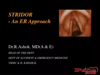
How to Approach and Manage Stridor. Children’s Hospital of New Orleans Louisiana State University Health Sciences Center, New Orleans. Lawrence M. Simon, M.D. Departmental of Lecture Series. Stridor. Harsh sound caused by turbulent airflow Implies partial airway obstruction

E N D
How to Approach and Manage Stridor Children’s Hospital of New Orleans Louisiana State University Health Sciences Center, New Orleans Lawrence M. Simon, M.D. Departmental of Lecture Series
Stridor • Harsh sound caused by turbulent airflow • Implies partial airway obstruction • Laryngeal stridor-inspiratory, biphasic
Congenital Laryngeal Anomalies Laryngomalacia-different types Tracheomalacia Vocal Cord Paralysis Laryngeal Clefts Vascular Rings and Slings Infectious “Croup” (Laryngotracheitis) Epiglottitis Tracheitis Trauma Croup Masquerade Subglottic Hemangioma Recurrent Respiratory Papillomatosis Post Intubation Glottic and Subglottic Lesions Congenital Glottic and Subglottic Stenosis Extra-Esophageal (Gastroesophageal) Reflux Disease/Eosinophilic Esophagitis Foreign Body Tracheal Esophageal Laryngeal Stridor: Etiology
Assessment Strategies • Guide to diagnosis and intervention • Age • Congenital vs. Acquired • Characteristics of stridor • Clinical picture
Clinical Picture: History • GERD symptoms • Wheezing episodes • Feeding problems: • FTT, weight gain • Choking episodes • Acute events • Onset: acute, chronic, progressive • Prior respiratory problems • Ex-premie (NICU stay) • Prior intubation
Clinical Picture: Associated signs & symptoms • Acute Disease • Fever • Drooling (new onset) • Change in cry or voice • Decrease in oral intake • Body position
Initial Evaluation • Assess urgency • Is there acute distress? • Nasal flaring • Tachypnea • Cyanosis • Retractions • Tripod position • Immediate action !
Physical Examination • Let parent hold child
Physical Examination • Let parent hold child • Mirror for nasal airflow (stertor vs stridor)
Physical Examination • Let parent hold child • Mirror for nasal airflow • “Headless” stethoscope
Physical Examination • Let parent hold child • Mirror for nasal airflow • “Headless” stethoscope • Positional changes
Pediatric Flexible Nasolaryngoscopy • Gold standard of office evaluation • Dynamic assessment • Easy to do • Minimal morbidity • Well tolerated
Pediatric Flexible Nasolaryngoscopy • Disadvantages • VC mobility often difficult to assess • Especially neonates • Excess secretions • Poor view of subglottis • Easily misinterpreted • Interpretation of erythema difficult • Overhanging epiglottis • Must be careful with local anesthesia in neurologically impaired child
Operative Endoscopy • Indications • To establish diagnosis or evaluate for synchronous lesions • Severe or progressive stridor • Cyanosis or apnea concerns • Radiologic abnormalities • Parental or physician anxiety • Anesthesiologist • Critical to success of operative evaluation • Comfort zone with sharing of the airway • Spontaneous ventilation
Concerns in Airway Endoscopy Postoperative edema, infection Long term treatment with steroids Extended hospitalizations, intensive care Complications: subglottic stenosis, glottic webs Voice abnormalities
Laryngomalacia • Most common cause of stridor in infants • Varying severity & varying types • Presentation • Staccato inspiratory stridor • Worse with exertion, feeding, crying • Noisy breathing generally begins at about 2-4 weeks of age • Office evaluation • Character of stridor • Positional changes / Mandibular thrust • Flexible nasopharyngolaryngoscopy
Laryngomalacia • Endoscopic appearance • Omega epiglottis • Foreshortenend aryepiglottic folds • Cuneiform prolapse Ω
Laryngomalacia • Vast majority are mild • Do these really need operative endoscopy? • Parental reassurance & education • Transient worsening, gradual improvement • Weight gain issues • GERD issues- Consider GERD treatment if there is evidence on endoscopy
Severe Laryngomalacia • Respiratory difficulty • Feeding difficulty • Failure to thrive • GERD • CNS abnormalities
Severe Laryngomalacia • Treatment • Aryepiglottic fold division (Aryepiglottoplasty) • Cold, Laser, Microdebrider • Treat presumed GERD
Dyscoordinate Pharyngolaryngomalacia • Absence of classical laryngomalacia findings • Prolapse of pharyngeal tissues • Neurologic abnormalities • Often older infants • Treatment Options: • Trial Bi-pap • Tracheostomy • Aryepiglottic fold division may make airway obstruction worse
Tracheomalacia • Symptoms/ Signs: • Tracheal wheeze • “Brassy” cough • Failure to thrive • Increasing respiratory distress with growth • Diagnosis: • Endoscopy – location, extent • May not be idiopathic- look for contributing factors • Treatment: • BiPAP / CPAP • Tracheotomy – variable tube length • Stenting – if no other choice
Croup (Laryngotracheobronchitis) • Begins about 6 months of age ! • “Croup” before 6 months is not croup • High KV AP of neck: symmetric subglottic narrowing (“steeple sign”) • Endoscopically: 2 “sets” of vocal cords • Hospitalized Patient: IV steroids, cold mist tent, hydration, O2 sat monitor
Epiglottitis • Traditionally caused by H. influenza b • Suden onset, rapidly progressive course • 80-90% decreased incidence since HIB vaccine introduced (1991) • Majority of cases seen now are caused by Staph • Consider immunocompromise • Treatment: • Immediate intubation in OR with ENT present • Send Cultures • Appropriate antibiotics
Tracheitis • Treatment: • Monitor, Humidified O2 • Bronchoscopy for suctioning of purulent secretions and culture • Antibiotics: • Consider Staph aureus (MRSA), H. flu, B-hemolytic strep, pneumococcus • Treat for 7-10 days • Tracheotomy in severe cases • Acute lower airway infection • Typically develops as bacterial super-infection after viral croup • Acute airway obstruction, high fever, elevated WBC develop 2-3 days after onset viral illness
Pediatric Airway Lesions Managed Endoscopically or with Open Surgical Repair • Subglottic hemangioma • Glottic and Subglottic stenosis, webs • Vocal Fold Immobility • Laryngeal Clefts • Saccular Cysts
Subglottic Hemangioma • Classic scenario • “Croup” symptoms generally begin 6-8 weeks of age • No fever, good cry • Respiratory distress/stridor: often treated as outpatient with oral steroids or inpatient with IV steroids with improvement • “Croup” recurs several weeks later • Mean age of diagnosis is 4 months • Delay due to misdiagnosis of symptoms • Proliferative phase then involutional phase
Subglottic Hemangioma • Assessment • Endoscope entire airway • Biopsy not essential (but can be done) • CT/MRI--r/o extraluminal extension
Subglottic Hemangioma • Classic Endoscopic appearance • Smooth submucosal mass • Posterolateral: left>right • Bilateral lesions mistaken as “soft” subglottic stenosis
Traditional Management Options for Airway Hemangioma Medical Steroids GERD therapy (Interferon-spastic diplegia concerns) (Vincristine-life threatening cases, neurotoxicity) Surgical Tracheostomy Open surgical excision +/- expansion LTR Endoscopic Excision CO2 / KTP laser Ablation Intralesional steroid injection
New Management Options for Airway Hemangioma Propanolol! Léauté-Labrèze C, Dumas de la Roque E, Hubiche T, Boralevi F, Thambo JB, TaïebA. Propranolol for severe hemangiomas of infancy. N Engl J Med. 2008 Jun12;358(24):2649-51.
Post Intubation Injuries • History • Intubation--even transient • NICU Graduate • Stridor with URI • History of recurrent or prolonged croup or asthma • Poor response to standard therapy
Post Intubation Injuries • Anterior commissure web • Weak, hoarse cry • Mild-moderate respiratory distress • Treatment: • Endoscopic division • Laryngeal keel • Short term post-op intubation • Mitomycin (?)
Posterior glottic injury: Progressive changes Post Intubation Injuries Granulation Ulceration Furrow Interarytenoid Scar
Interarytenoid web Difficult problem Mistaken for vocal cord paralysis Assess with MSL using 2 handed distraction technique Repair: endoscopic division alone rarely successful Mitomicin C Mucosal flap interposition Posterior cricoid expansion Post Intubation Injuries
Post Intubation Injuries • Subglottic Stenosis • Assess entire airway • Size & grade stenosis • Grade I (0-50%) • Grade II (50-70%) • Grade III (70-99%) • Grade IV (No visible lumen) • Treatment • Observation (Grade I-II) • Dilatation (Grade II-III) • Laryngotracheal reconstruction (Grade III-IV) • Treat for GERD
Post Intubation Injuries • Subglottic Cysts • Often multiple • Removal • Forceps • Laser (CO2 / KTP) • Microdebrider