Download

1 / 46
460 likes | 762 Views
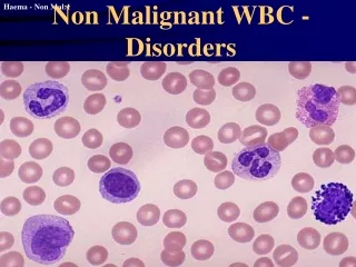
The Non- leukaemic Malignant Disorders . Ahmad Sh. Silmi Msc Haematology, FIBMS. Definition .

E N D
The Non-leukaemic Malignant Disorders Ahmad Sh. Silmi Msc Haematology, FIBMS
Definition • The Non-leukaemic Malignant Disorders are originally defined, as an amorphous group of malignant disorders which are characterized by the uncontrolled clonal proliferation of bone marrow derived cells.
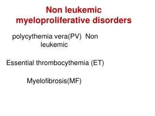
Classification • 1. The malignant myeloproliferative disorders, which include: • Primary proliferative polycythaemia (PPP) • Primary thrombocythaemia • Myelofibrosis • 2. The malignant lymphoproliferative disorders, which include: • Multiple myeloma (MM) and related plasma cell disorders. • Hodgkin's disease (HD) • Non-Hodgkin's lymphomas
Introduction • Hemopoietic stem cell disorder • Clonal • Characterized by proliferation • Granulocytic • Erythroid • Megakaryocytic • Interrelationship between • Polycythaemia • Essential thrombocythaemia • myelofibrosis
Clonal evolutionClonal evolution & stepwise progression to fibrosis, marrow failure or acute blast phase
1- Primary Proliferative Polycythaemia • Primary proliferative polycythaemia is a malignant disorder of haemopoietic stem cells, which is characterized by absolute erythrocytosis and, a moderately increased granulocyte count and platelet count. • It may occur at any age, but is predominantly a disease of middle age: the median age at diagnosis is 55 years. Males are affected slightly more commonly than female.
Polycythaemia vera(Polycythaemia rubra vera) • Polycythaemia vera is a clonal stem cell disorder characterised by increased red cell production • Abnormal clones behave autonomous • Same abnormal stem cell give rise to granulocytes and platelets • Disease phase • Proliferative phase • “Spent” post-polycythaemic phase • Rarely transformed into acute leukemia
Recognition and classification of the polycythaemia • Definition of polycythemia • Raised packed cell volume (PCV / HCT) • Male > 0.51 (50%) • Female > 0.48 (48%) • Alterations in the PCV can be caused by: • Reduction in plasma volume. • True increase in red cell numbers.
Classification • Absolute • Primary proliferative polycythaemia (polycythaemia vera) • Secondary polycythaemia • Idiopathic erythrocytosis • Apparent • Plasma volume or red cell mass changes
Polycythemia • True / Absolute • Primary Polycythemia • Secondary Polycythemia • Epo dependent • Hypoxia dependent • Hypoxia independent • Epo independent • Apparent / Relative • Reduction in plasma volume
Absolute Polycythaemia • Causes: • Malignant transformation, which frees the haemopoietic stem cells from hormonal control. • Hypoxic effect. • Physiologically inappropriate stimulation of erythropoietin release. • Thus the polycythaemias are large and diverse group of disorders, only one member of which, PPP, is a malignant disorder. However, an understanding of all types of polycythaemia is essential because recognition of PPP largely is based upon exclusion of the other types.
Causes of secondary polycythemia • ERYTHROPOIETIN (EPO)-MEDIATED • Hypoxia-Driven • Chronic lung disease • Right-to-left cardiopulmonary vascular shunts • High-altitude habitat • Chronic carbon monoxide exposure (e.g., smoking) • Hypoventilation syndromes including sleep apnea • Renal artery stenosis or an equivalent renal pathology • Hypoxia-Independent (Pathologic EPO Production) • Malignant tumors • Hepatocellular carcinoma • Renal cell cancer • Cerebellar hemangioblastoma • Nonmalignant conditions • Uterine leiomyomas • Renal cysts • Postrenal transplantation • Adrenal tumors • EPO RECEPTOR–MEDIATED • Activating mutation of the erythropoietin receptor • DRUG-ASSOCIATED • EPO Doping • Treatment with Androgen Preparations
Secondary polycythaemia • Polycythaemia due to known causes • Compensatory increased in EPO • High altitude • Pulmonary diseases • Heart dzs eg- cyanotic heart disease • Abnormal hemoglobin- High affinity Hb • Heavy cigarette smoker • Inappropriate EPO production • Renal disease-carcinoma, hydronephrosis • Tumors-fibromyoma and liver carcinoma
Secondary Polycythaemias • Causes: • Physiologically appropriate release of erythropoietin. • Physiologically inappropriate release of erythropoietin.
1- Physiologically appropriate release of erythropoietin. • A persistently low atmospheric oxygen tension: • As in high altitude • Inadequate uptake of atmospheric oxygen: as in • chronic bronchitis • emphysema • pulmonary fibrosis, • pulmonary oedema • Defective transport of absorbed oxygen from the lungs:This occurs in the following abnormalities: • A variety of inherited and acquired defects of haemoglobin function. • Metabolic defects, which limit the effectiveness of oxygen transport, include methaemoglobin reductase deficiency and 2,3 DPG deficiency. • Heavy smoking, which affect the haemoglobin function.
2- Physiologically inappropriate release of erythropoietin • A variety of disorders are associated with inappropriate release of erythropoietin occurs as a rare complication of renal disorders such as: • polycystic kidneys. • chronic glumerulonephritis. • Hydronephrosis. • renal tumours. • extrarenal tumours such as hepatoma and bronchial carcinoma.
Secondary polycythaemia • Arterial blood gas • Hb electrophoresis • Oxygen dissociation curve • EPO level • Ultrasound abdomen • Chest X ray • Total red cell volume(51Cr) • Total plasma volume(125 I-albumin)
Apparent Polycythaemia • Apparent polycythaemia is defined as those cases where an increased venous PCV is not explained by an increased red cell mass (RCM) but by a reduction in the plasma volume (PV), or by the combination of an RCM towards the upper limit of normality and a PV towards the lower limit of normality. • Apparent polycythaemia is by far the most common finding where the PCV is only minimally increased, accounting for up to 50% of such cases. • Alternative names for this condition include relative polycythaemia, pseudopolycythaemia, stress polycythaemia and spurious polycythaemia.
Apparent Polycythaemia • Causes • Stress • Cigarette smoker or alcohol intake • Dehydration • Plasma loss- burn injury
Pathophysiology • The presenting features of PPP are related to hypervolaemia and hyperviscosity, which accompanies the absolute increase in red cell mass. Typical complaints include: • Headache • Blurred vision • Dizziness • Mental impairment • Feeling of congestion on the head
Pathophysiology • Splenomegaly secondary to extramedullary haemopoiesis. • Hyperviscosity with impairment of blood flow contribute to the increased incidence of arterial and venous thrombosis. • Platelet dysfunction with a tendency to bruise and bleeding tendency. • Sever itching particularly after a warm bath, due to histamine release from basophile.
Laboratory findings-1 • Increase Hb, PCV and RBCs • Increase granulocytes count • Neutrophil morphology and function are increased • Platelet increases by about 50%, by bizarre platelet morphology and dysfunction are common. • Bone marrow is hypercellular with predominance of granulocytes. Erythroid hyperplasia is less common. • Cytogenetic abnormalities are present in about 15% of cases at presentation.
Laboratory findings-2 Bone marrow in PV • Normal Neutrophil Alkaline Phosphatase (NAP). • Plasma urate high • Circulation erythroid precursors • Low serum erythropoietin
Clinical features • Majority patients present due to vascular complications: • Thrombosis (including portal and splenic vein) • DVT • Hypertension • Headache, poor vision and dizziness • Skin complications (pruritus, erythromelalgia) • Haemorrhage (GIT) due to platelet defect
Polycythaemia vera(Polycythaemia rubra vera) Erythromelalgia • Hepatosplenomegaly • Erythromelalgia • Increased skin temp • Burning sensation • Redness Liver 40% Spleen 70%
Prognosis • 25% of PPP transform to chronic myelofibrosis and only 1/3 of these progresses to ANLL. The treatment with chemotherapy is associated with increase transformation to ANLL.
Treatment • The main aim is to reduce thrombotic complication. • " Debulking" by removal of RBCs and platelet by apheresis or by repeated phlebotomy. The median survival may be as long as 15 years. • Additional treatment may not be needed and is controversial(Chemotherapy).
2- Primary Thrombocythaemia • Primary thrombocythaemia is a malignant clonal disorder, which is characterized by megakaryocytic hyperplasia and a markedly increased circulating platelet count. • Alternative names for this disorder include essential thrombocythaemia, idiopathic thrombocythaemia and primary haemorrhagic thrombocythaemia.
Incidence • Rare in children and young adult. • Median age is 60 years. • Men and women appear to be affected with equal frequency.
Pathophysiology • Increase thromboembolism due to increase in platelet count. • Increase haemorrhage due to platelet dysfunction. • Splenomegaly in more than 80% of patients. • Some hepatomegaly.
Clinical Features • Increase platelet count for up to 10X106/ m3. • Platelet clumping with bizarre shape. • Marked platelet variation. • Fragment megakaryocytes. • Increase WBC. • Normal Hb and RBCs. • Iron deficiency after chronic haemorrhage. • 25% transfer to myelofibrosis or PPP or ANLL.
Treatment • Many do not require. • Platelet apheresis. • Alkalating agent such as melphelan or busulphan. • Heparin or Warfarin. • Aspirin as a prophylactic.
Prognosis • Median survival time is 10 years. • Disease course and prognosis • 25 % develops myelofibrosis • Acute leukemia transformation • Death due to cardiovascular complication
3- Myelofibrosis • Myelofibrosis predominantly is a disease of the middle-aged and elderly. • It's characterized by: • Progressive collagen fibrosis of the bone marrow. • Megakaryocytic hyperplasia. • Splenomegaly due to extramedullary haemopoiesis. • Myelofibrosis affects men and women equally.
Pathophysiology • Lethargy and exercise intolerance secondary to anaemia. • Weight loss and night sweats secondary to metabolic disturbances. • Bruising secondary to platelet dysfunction. • A feeling of left-sided abdominal fullness secondary to massive splenomegaly secondary to extramedullary haemopoiesis. • Extramedullary haemopoiesis in liver may be present. • Greatly increased bone density with narrowing of the medullary cavity.
Laboratory Findings • Peripheral blood: • Leukoerythroblastic anaemia with prominent polychromasia, anisocytosis and "tear drop" poikilocytosis. • The WBC and platelet count are variable but frequently are raised. • As the disease progresses: • The degree of dysplasia increases. • The circulating cell counts markedly decreased. • Increase WBC and RBC turnover leading to increase LDH, uric acid and lysozymes and in advanced cases, the serum concentrations of hepatic enzymes may be abnormal.
Leucoerythroblastic blood film • Tear drops red cells • Increase in NAP score
Laboratory Findings • Bone Marrow: • Failure to aspirate bone marrow because of the fibrotic overgrowth of marrow space. • Bone marrow smear reveals large patchy areas of hypercellularity, which contain prominent clusters of dysplastic megakaryocytes. • These megakaryocytes die within the marrow space and release platelet-derived growth factor (PDGF) and platelet factor 4 (PF4). • PDGF is a fibroblast mitogen. • PF4 inhibits neutrophil and fibroblast collagenase enzymes. The combined effects of these two substances rise in collagenous fibrosis of the marrow, which characterized this condition.
Bone Marrow, cont. • Marrow fibrosis thus is a secondary phenomenon in myelofibrosis, the primary defect is thought to lie in a haemopoietic progenitor cell of CFU-MK and CFU-GM. • The increasing hostility of the marrow haemopoietic microenvironment promotes the establishment of extramedullary haemopoiesis in the foetal sites of the spleen and liver.
Treatment and Prognosis • The median survival time is 3 years but some cases survive for more than 10 years. • About 10 % of cases progress to ANLL mostly in men. • Splenic irradiation to reduce the size of the spleen and to relieve the symptoms of massive splenomegaly such as red cell or platelet sequestration. Where irradiation is ineffective splenectomy may be performed. • Blood transfusion when required to correct anaemia. • Androgen to correct ineffective haemopoiesis. • Haematinic therapy if iron or folate deficiency is presents. • Allupurinol to correct hyperuricaemia. • Where the disease is advancing rapidly, chemotherapy can be used with limited success. • The only hope is bone marrow transplantation in young ages.