Download

1 / 11
110 likes | 172 Views
Effects of THz exposure on Human Primary Keratinocyte Differentiation and Viability. Clothier, R.H. , and Bourne, N. School of Biomedical Sciences, Faculty of Medicine and Health Sciences, University of Nottingham, Nottingham. NG7 2UH, UK.

E N D
Effects of THz exposure on Human Primary Keratinocyte Differentiation and Viability. Clothier, R.H., and Bourne, N. School of Biomedical Sciences, Faculty of Medicine and Health Sciences, University of Nottingham, Nottingham. NG7 2UH, UK.
Effects of THz exposure on Human Primary Keratinocyte Differentiation and Viability. Primary human keratinocytes driven, in vitro, to differentiate via activation of transglutaminases, results in transglutaminase regulated cross linking of specific amino acids with cornified envelope formation monitored via the incorporation of fluorescein cadaverine, as a substitute for L-Lysine (Gray et al., 1999). This differentiation endpoint was co-assayed with the keratinocytes reductive capacity for converting resazurin to resorufin, as a measure of cell activity/viability (Andrew et al., 1997; Rasmussen 1999; O’Brien et al., 2002; Gray and Clothier 2001).
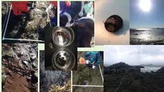
Effects of THz exposure on Human Primary Keratinocyte Differentiation and Viability. • Materials and Methods • Primary Human Keratinocytes isolated from human skin, donated with patient consent. The primary keratinocytes were passaged once, then the cells were seeded (passage 2) from a 5x10⁴/ml suspension, at 100µl per well into a 96 well plate. • The plates were placed into a thermal box at room temperature (approx. 22°C) and transported to the THz source in HBSS. • THz Exposure • This took place at TeraView Ltd, Cambridge, or The Institute of Microwaves and Photonics, University of Leeds. The keratinocytes were exposed to the THz through the base of the 96 well plate in clusters of 4 wells per exposure time.These both used a Ti:Sapphire laser impacting on an electro-optic photoconverter to generate THz power. The Leeds system had a total pulse duration is 20-30ps, (90% delivered within two ps). The average output power for this (unamplified) system is approximately 1W within the frequency range 0.2 – 3.0THz. The repetition rate of the THZ pulse is approximately 80MHz . • At Teraview by optical excitation of a gallium arsenide wide aperture antenna. A large DC-bias applied across the device which was excited using a Ti:Sapphire laser emitting 250 fs pulses centered at a wavelength of 800 nm, with a 250kHz repetition rate. Frequency range of 0.1 THz to 2.7 THz, average power of approximately 1 mW. The THz-radiation was focused paraobla mirror on to the sample with a spot size of 130 µm to 3.7 mm. The THz spot was raster scanned over sample for the duration of the exposure.
Effects of THz exposure on Human Primary Keratinocyte Differentiation and Viability. Materials and Methods Cont. Prior to the exposure to the THz, a resazurin (Sigma, Poole, Dorset) assay was performed. Cells were exposed for periods of 10, 20 or 30 minutes, giving a total exposure of 0.15, 0.3 or 0.45mJ/cm2 or 0.15, 0.3 or 0.45J/cm2 per 4 wells. Two sets of 4 control wells were exposed to the same conditions but not the THz beam. The plates were returned to the thermal box and transported back (2 hours). A resazurin assay was then performed and again at 3, 6 and 8 days. Following the initial post exposure resazurin assay the keratinocytes were returned to Greens Medium (Gray et al., 1999) containing 20µM/ml fluorescein cadaverine (Sigma; Gray et al., 1999; Gray & Clothier., 2001) to stimulate and quantify differentiation.
Effects of THz exposure on Human Primary Keratinocyte Differentiation and Viability. Methods Grow to confluence Isolate Human Keratinocytes Seed to 96 well plates, 5000 per well Perform Resazurin assay Return at 23 C & transport to Nottingham (2 hrs) THz Exposure for 10, 20 or 30 mins Place in HBSS at 23 C & transport to THz source (2 hrs) Resazurin assay at 3 days then into Green’s Medium with 20µm Fluorescein Cadaverine Resazurin assay 6 days post exposure Resazurin and FC assayed (return to Greens Medium with 20µm FC) 8 days post exposure Resazurin and FC assayed Fix cells.
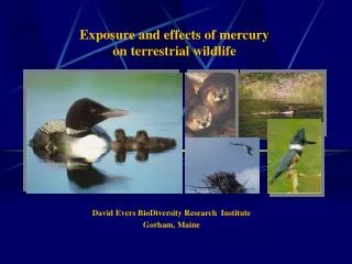
Effects of THz exposure on Human Primary Keratinocyte Differentiation and Viability. Typical coblestone appearance of keratinocytes. Fluorescein Cadaverine assays differentiated cells green Resazurin assay with cells converting it to resorufin being red. Confocal image.
Effects of THz exposure on Human Primary Keratinocyte Differentiation and Viability.
Effects of THz exposure on Human Primary Keratinocyte Differentiation and Viability.
Effects of THz exposure on Human Primary Keratinocyte Differentiation and Viability.
Effects of THz exposure on Human Primary Keratinocyte Differentiation and Viability. • The THz from the two sources gave comparable results with no signs of induced stress to the cells, over the subsequent culture period. • From previous results and the database on FC incorporation rates, it has been shown that there is a rise in FC uptake in normal keratinocytes from <20pg per well to 111 ± 38pg per well on 3 and 9 days respectively. • The results presented, with an average of 184 ± 85pg per well were in line with these results. • The differentiation capacity was as expected for all three donors, exposed to the higher THz levels up to 0.45J/cm2. This was also true for the donors exposed to the lower energy levels. • Thus the THz caused not detectable change in the activity or differentiation capacity of confluent primary human keratinocytes from the basal and stem cell population of the stratum basale of the epidermis. • Dividing cells will need to be examined.
Effects of THz exposure on Human Primary Keratinocyte Differentiation and Viability. We acknowledge the funding under the FW5 programme THz-Bridge (QOL-2000-4.2.2), and the assistance of the FRAME Research Laboratory staff, THz generation under the control of Emma Pickwell, Cambridge University or Vincent Wallace and Philip Taday, Teraview, Cambridge Science Park, Cambridge, or Dr. M.A.Naftaly and Prof. N.N.Zinovev, Teravision, ( Institute of Microwaves & Photonics), School of Electronics & electrical Engineering, The University of Leeds, Leeds LS2 9JT (funded under the EPSRC and by the EU as part of the Teravision programme (IST-1999-10154).