Download
1 / 24
270 likes | 488 Views
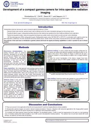
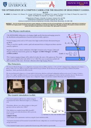
Development of a camera for imaging of prompt gamma rays in measurements of proton beam range. P.Busca 1,2 , C.Fiorini 1,2 , R.Peloso 1,2 , M.Basilavecchia 3 , T.Frizzi 3 , D. Prieels 4 , J. Smeets 5 * , F. Stichelbaut 4 , A. Benilov 4 , F. Roellinghoff 4

E N D
Development of a camera for imaging of prompt gamma rays in measurements of proton beam range P.Busca1,2, C.Fiorini1,2, R.Peloso1,2, M.Basilavecchia3, T.Frizzi3, D. Prieels4, J. Smeets5*, F. Stichelbaut4, A. Benilov4, F. Roellinghoff4 1Politecnico di Milano and INFN, Milano, Italy 2XGLab, Milano, Italy 4 Ion Beam Applications S.A., Louvain-la-Neuve, Belgium 5 Department of Nuclear Metrology, UniversitéLibre de Bruxelles, Brussels, Belgium • Work supported in part by the EuropeanUnionSeventhFrameworkProgram (FP7/2007-2013) under grantagreementsnos. 241851 (ENVISION) and 264552 (ENTERVISION). • HICAM gamma detector developedwithin the EC contract n.LSHC-CT-2006-037737. • *supported by the Belgian FNRS (aspirant)
Proton therapy Bragg-peak • particle (proton) therapy has a growing role in cancer treatment. • possibility to release the maximum of the dose in the target site, limiting the dose to normal tissue relative dose g p depth • the measurement of the proton beam range in the target is very important: real range of proton beams in patients may contain uncertainties of up to 10-15 mm (uncertainties on tissue composition, density, organ motions, patient positioning, etc).
One method to measure proton beam range is based on the measurement of prompt gamma rays (energies up to 10MeV) emitted by excited nuclei during proton irradiation. (F. Stichelbaut, Y. Jongen, 39th Meet. of the ParticleTherapy Co-Operative Group, San Francisco, October 2003) p beam prompt gs p beam target Monte Carlo simulation
Practical concept of a prompt gamma camera which allows checking in real-time the range of a single pencil beam with a ‘mm’ accuracy (D. Prieels, J. Smeets, F. Stichelbaut, A. Benilov, J.C. Dehaes, A. Dubus, F. Roellinghoff, 50th Meet. of the ParticleTherapy Co-Operative Group (Philadelphia), May 2011
HICAM:HIgh resolution gamma CAMera Researchproject fromEuropeanCommunity (www.hi-cam.org) Anger camera principle drift scintillation entering side Silicon Drift Detectors • - CsI(Tl) scintillator • - SDDs photodetectors • - 5x6cm2 and 10x12cm2 formats • intrinsic resolution~1mm • compactness
The HICAM gamma camera development • (R.Peloso, et al., “The HICAM Gamma Camera”, Nuclear Science Symposium Conference Record (NSS/MIC), 2010 IEEE)
Imaging performances with 99Tc (140keV) Grid 8,7 cm intrinsic resolution= 0.8mm Hole Ø=1mm Pitch=3mm corrected for linearity and uniformity LEUHR parallel hole collimator 10,8 cm 99Tc
Applicationsof the HICAM gamma camera (P.Busca, et al., “Applications of the HICAM Gamma Camera”,Nuclear Science Symposium Conference Record (NSS/MIC), 2010 IEEE)
Modificationof the 5x6cm2 HICAM gamma camera forhigh-energy (1-10MeV) imaging • 1cm thick LYSO scintillator • (h~37%@1MeV, ~22%@5MeV) • light collection purposely limited (i.e. all absorbing surfaces) to match readout ASIC dynamic range (designed for 200keV) • ( ~ 5e-/keV, vs. 30ph./keV from LYSO)
Laboratorycharacterizationof the camera 1D profilecalculationalong X: integration on the Y direction (1mm bin) Source: 60Co (1.17MeV, 1.33MeV) Y Camera FOV source lead block X Area consideredforprofilecalculation lead block thickness=3cm
Proton range measurements: the experimental set-up p beam slitcollimator opening: 6mm
electronics HICAM camera g-rays proton beam slit collimator PMMA target
Measured energy spectrum LYSO background 3-7MeV range used in reconstruction
Measured profiles Beamenergy: 160MeV slits closed (neutrons + uncorrelated photons) camera position + 4cm camera position - 4cm camera centered subtraction central profile is fitted and monitored while moving the target (and the Bragg-peak) simulation
100MeV protons (preliminary analysis) -10mm +10mm • each acquisition: • 5min. • 7·1010 protons
Shift accuracy reference value X10 X0 X-10
Precision reference value reference position fitting error
Precision vs. statistics 7·1010 protons
160MeV protons (preliminary analysis) • each acquisition: • 2min. • 1.7·1010 protons
Conclusions • Measurements of prompt gammas correlated to proton range were successfully made using the HICAM gamma camera • Although not optimized for 1-10MeV energy range (low efficiency, low scintillator light collection), satisfactory accuracy and precision (in the ‘mm’ range) were first measured Future work • Further data analysis also to correlate results to dose expected in patient treatment. • Measurements to be repeated also with other protons energies (230MeV). • Gamma camera to be modified/redesigned to improve efficiency and speed