Download

1 / 32
340 likes | 670 Views
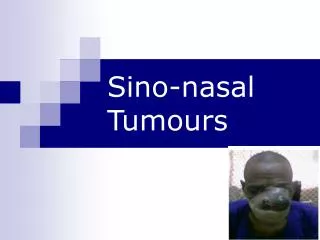
Sino-nasal Tumours. Tumors of the nasal cavity proper are approximately evenly divided between benign and malignant neoplasia , with inverting papilloma predominating in the benign group and squamous cell carcinoma in the malignant.

E N D
Tumors of the nasal cavity proper are approximately evenly divided between benign and malignant neoplasia, with inverting papilloma predominating in the benign group and squamous cell carcinoma in the malignant.
On the other hand, most sinus tumors are malignant with squamous cell carcinoma being the most prevalent. • The maxillary sinus is most commonly involved with tumor, followed by the nasal cavity, the ethmoids, and then the frontal and sphenoid sinuses.
Inverted papillomas • Their etiology is unclear. • They are known to have high recurrence rates. • They are associated with malignancy and also have locally aggressive growth patterns, which makes them technically difficult to remove. • There is also controversy over the appropriate surgical approach for tumor removal. • There is a role for radiation therapy.
Inverted papillomas • Papillomas differ from inflammatory polyps, which are more common, in that inflammatory polyps are associated with allergic rhinitis and are actually reactive lesions, not a tumor. • Nasal papillomas are true neoplasms and, while their etiology is unclear, they are known to arise from the nasal respiratory epithelium, which undergoes metaplastic change and proliferation.
Inverted papillomas arise from the Schneiderian membrane, which is an invagination of the olfactory ectoderm that occurs during the fourth week of embryonic development. The mucosa creates a transitional zone between the endodermally-derived respiratory epithelium of the nasopharynx and keratinizing squamous epithelium with the nasal vestibule. • Three types: Fungiform papillomas, cylindrical papillomas and inverted papillomas.
Grossly, inverted papillomas appear more opaque than inflammatory polyps, and this is because they have a thick epithelial layer. • Inverting papilloma traces its name to the histologic appearance with squamous epithelium inverted in the polyps
They are commonly located in the nasal cavity and they typically involve an adjacent sinus. • The most common location is the middle turbinate, but other common locations include the ethmoid sinus and maxillary sinus. • They have even been found in the nasopharynx.
Most common symptoms are unilateral and include nasal obstruction, nosebleed and nasal discharge. It can be an incidental finding on examination. • These tumors are rare. They occur about 0.6 cases per 100,000 per year and they occur approximately 1/25th as often as inflammatory polyps. • The average age at diagnosis is 53, but can range anywhere from the pediatric age of 6 to the elderly age of 89. • They are known to have a male predominance of (1:3) • The recurrence rates cited in the literature varies anywhere from 11% to 78%, and this depends a lot on the treatment modality used.
They are associated with malignancy, 5% to 15% malignancy rates are most generally accepted. Inverted papillomas are more commonly associated with squamous cell carcinomas. • There are four types of association: • Metachronous squamous cell carcinoma. • Carcinoma in situ within the IP • Synchronous lesions • Malignant transformation
The mainstay of treatment is surgery, although radiation therapy can be involved. • Traditionally procedures have been either a transnasal procedure with polypectomy or confined transnasal polypectomy with additional sinus procedure, such as Caldwell-Luc. The gold standard was lateral rhinotomy with medial maxillectomy. • Radiation therapy can be used as the sole therapy for inverted papilloma or it can be used postoperatively. The absolute indication for radiation therapy is when an inverted papilloma is associated with squamous cell carcinoma • The patients who should get radiation therapy are those who had advanced incompletely resected or unresectable lesions that are biologically aggressive, or patients where morbidity in resection would be more pronounced that morbidity of tumor radiation.
Sinonasal neoplasms • These are rare, comprising less than 3% of all malignant aerodigestive tumors and less than 1% of all malignancies. • Despite their infrequence, they represent both a diagnostic and therapeutic challenge because the presenting signs and symptoms may be indistinguishable from benign or inflammatory disorders. • These malignancies typically affect Caucasion males in the fifth to seventh decades of life and have a 2:1 male preponderance.
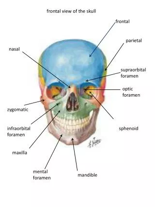
Sinus Anatomy: Maxillary antrum Significance • Superior – orbit, ethmoids • Posterior – pterygoids, infratemporal fossa • Ethmoid sinus Significance • Superior – fovea, cribiform • Medial – lamina papyracea
Sinus Anatomy: • Sphenoid sinus Significance • Superior – optic nerve, pituitary • Lateral – ICA, cavernous sinus • Lateral wall < 0.5mm • Inferior – NP, vidian nerve
Frontal sinus Significance • Inferior – orbit • Posterior – anterior cranial fossa
Lymphatic Drainage • The anterior nose has the same lymphatic drainage as the external nose. These tend to spread to the submental or level I area. • The posterior nose tends to drain to the retropharyngeal nodes as well as the lateral pharyngeal nodes, which eventually drain into the level II.
Epidemiology • Despite this, up to 44% are attributed to occupational exposures, including nickel, chromium, isopropyl oils, volatile hydrocarbons, and organic fibers that are found in the wood, shoe, and textile industries. • In addition, human papilloma virus can be a cofactor, and in one series, human papillomavirus 6 or 12 was documented in 24% of inverting papillomas and 4% of squamous cell carcinomas. • Specific asssociations found include squamous cell carcinoma in nickel workers and adenocarcinoma in workers exposed to hardwood dusts and leather tanning.
The most common entities are squamous cell carcinoma. The lateral nasal wall is the most common site of involvement, but SCC can also present in the sinuses. • Regional lymph node metastasis is more common with squamous cell than most other paranasal sinus malignancies, occurring in about about 10% to 20%. Local recurrence rates are quite high, as high as 30% to 40% • Adenocarcinoma is the second most common malignancy in this area. It is most often in the ethmoids, has a male predominance, and is often seen in industrial workers.
About 3% to 15% of these paranasal sinus malignancies are adenoid cystic carcinoma. It is occurs most frequently in women, and in the fifth and sixth decades. • Melanoma is rarely seen, comprising only about 3% of these paranasal sinus malignancies. • Olfactory neuroblastoma or esthesioneuroblastoma are neural crest in origin, and they arise from an olfactory epithelium.
Signs and symptoms of maxillary sinus carcinoma fall into several major categories • Oral • Nasal • Ocular, facial • Auditory
Oral presentations occur in 25-35% and include pain involving the maxillary dentition, trismus, palatal and alveolar ridge fullness, and frank erosion into the oral cavity. • Nasal findings are seen in up to 50% of patients and include obstruction, discharge, stuffiness, congestion, epistaxis, and extension into the nasal cavity.
Ocular findings occur in approximately 25% and arise from upward extension into the orbit, where unilateral tearing, diplopia, fullness of lids, pain, and exophthalmos are seen
Facial signs include infra-orbital nerve hypoesthesia, cheek swelling, pain, and facial asymmetry. • Auditory complaints include hearing loss secondary to serous otitis media due to nasopharyngeal extension. • With advanced disease, the classic triad of findings for carcinoma of the nasal cavity and paranasal sinuses may be present: These include • Facial asymmetry • A visible or palpable tumor bulge in the oral cavity • Tumor visible in the nose with anterior rhinoscopy.
Staging: • Ohngren line, a line that is drawn from the angle of mandible to the medial canthus. Ohngren indicated that tumors that presented above this line, both superiorly and posteriorly, tended to have a worse prognosis
American Joint Committee on Cancer Staging System is the gold standard used for reporting in most professional papers. • T1 tumors of the nose and nasal cavity, and ethmoid sinuses, are tumors restricted to any one sub-site, with or without bony invasion. • T2s are tumors invading two sub-sites, single region or extending to involve adjacent regions of the nasal ethmoid complex. • T3 tumors begin to have bony involvement, invading the medial wall of the floor of the orbit, cribriform or palate. • T4-A tumors involve the anterior orbital contents, nose, and cheeks, with extension into the anterior cranial fossa. • T4-B, involve the orbital apex, dura, middle cranial fossa, as well as the clivus.
Investigations • CT scans are excellent for determining bony erosion and extent of invasion.
If there is a question of neural involvement, MRI is excellent for determining perineural spread, involvement of the dura, or involvement intracranially.
Lastly, confirm diagnosis via biopsy. Most often biopsy is performed after imaging rule out encephaloceles or other vascular issues. • PET scan has been used to evaluate for residual tumor, recurrent tumor, and radiated treated fields. • Angiography is not initially used, but can be used for vascular tumors to determine extent and vascularity as well as to allow for embolization prior to any surgical interventions.
Management • Adenocarcinoma: Treatment is controversial, but the literature indicates that craniofacial resection is the key.
Management • SCC: For the treatment of early lesions, surgery, if the tumor is excised en bloc with good margins, and, if there is no evidence of perineural spread, then surgery is usually sufficient. If there are any questions about the margins or perineural invasion, the addition of radiation is indicated External Inferior medial Medial Radical
Management • There has been some literature reporting the use of radiation therapy alone for early disease, but this is not necessarily recommended since radiation of this side of the body has significant morbidity, with possible osteoradionecrosis and vision loss as well as damage to the spinal cord. • Combined modality generally tends to be the gold standard: surgery with postoperative radiation therapy.
Management • Also, the use of chemotherapy is now being added with the goal of better local control and improvement in survival. • Chemotherapy does have a role in palliation for large tumors that are nonresectable. • If there is nodal disease of the neck with squamous cell carcinoma, a neck dissection is generally indicated