Download
1 / 35
840 likes | 2.7k Views
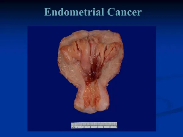
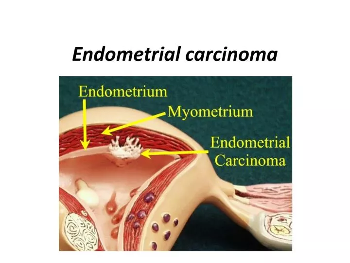
Endometrial carcinoma. Endometrial carcinoma is the fifth leading cancer in the women worldwide. In developed countries it’s the most common gynaecological cancer but in developing countries it’s surpassed by cervical cancer. Age groups: Mean age of presentation is 56 years .

E N D
Endometrialcarcinoma is the fifth leading cancer in the women worldwide. In developed countries it’s the most common gynaecological cancer but in developing countries it’s surpassed by cervical cancer.
Age groups: Mean age of presentation is 56 years . 75% after menopause. 20% perimenopausal. 5% before age of 40. Aetiology: a- indiscriminate use of oestrogen. b- un opposed oestrogen. c- Theca granulosa cell tumours.
Prediposing factors: Atypical adenomatous hyperplasia (complex atypical ) –25% (10 - 60%) to progress to cancer Type of patient: Nullipara or low parity Middle or upper social class Overweight and obese patients Early menarche and late menopause
Associated factors: • Diabetes or abnormal glucose tolerance test. • Hypertension. • Fibroids. • Polycystic ovarian syndrome. • Infertility, Arthritis, and Thyroid disease. • Use of TAMOXIFEN. • Previous pelvic irradiation. • Positive family history of breast, ovarian, and to lesser extent colon cancer.
Protective factors: Smoking ! Use of oral contraceptive. Use of progesterone.
Pathology: Adenocarcinoma ------------------------------------------------59% Adenoacanthoma(adeno + squamous metaplasia)------21% Adenosquamous carcinoma------------------------------------7% Clear cell carcinoma----------------------------------------------6% Papillary adenocarcinoma---------------------------------------5% Secretory carcinoma----------------------------------------------2% Mixed type------------------------------------------------------------ ~
Spread : • Invasion through the myometrium and by filling the uterine cavity. • Invasion to the cervix with subsequent lymphatic spread involving the iliac and para-aortic nodes. • From upper uterus may spread to round ligament to the deep inguinal nodes. • In advanced cases, the blood-stream spread may carry to the lungs, liver, and to the bone.
In general 95% adenocarcinoma and 5% squamous cell carcinoma. • More often well differentiated than anaplstic. • May be associated with pyometra or haematometra secondary to cervical stenosis.
Diagnosis and assessement: A-History: • postmenopausal bleeding or staining( this symptom should be assumed to be caused by carcinoma of the endometrium until proved otherwise), only 10% of PMB have endometrial carcinoma. • Perimenopausal menstrual irregularities. • Blood stained vaginal discharge. • Heavy and irregular vaginal bleeding.
Diagnosis and assessement: B-Examination: • physical examination of the patient with endometrial carcinoma is frequently entirely normal, it should include palpation of supraclavicular and inguinal lymph nodes, abdominal palpation might be difficult due to obesity. • Gynaecological examination: • inspection of vulva, vaginal skin in suburethral area • and cervix. • Bimanual vaginal examination assesses uterine size, and mobility, state of parametria and adnexa. • Bimanual recto-vaginal examination.
Diagnosis and assessement: C-Investigation: • CBC. • Liver function test. • Renal function test. • Chest X ray. • Cytology brush from lower cervical canal and posterior fornix to analyze the cells. • Endometrial sampling :-
Endometrial sampling :- ONE sample for histology: - Piplle - Vabra - Jet suction - Other Two Examination under anaesthesia and D&C. Three Hysteroscopy & biopsy. -ultrasound for endometrial thickness, myometrial invasion and lymph nodes. -MRI – to assess site, thickness, and myomtrial invasion for staging. -proctoscopy and / or sigmoidoscopy, cystoscopy, and bone scan for some exceptional cases, when there is a clinical suspicion of metastasis.
Staging : • The staging was changed in 1988 from a clinical staging to a surgical staging which is far more realistic and accurate. • The initial examination is clinical, aimed to take biopsy to diagnose and record an initial staging to facilitate planning of the subsequent surgical procedure. The clinical staging used for advanced stage to be treated with radiotherapy. The staging is only finalized after histopathologic examination of surgically removed tissue.
Stage I: Carcinoma confined to the corpus I a – tumour limited to endometrium. I b – invasion of less than ½ of myometrium. I c – invasion of more than ½ of myometrium.
Stage II: Extension to the cervix. II a – Endocervical glandular involvement. II b – Cervical stromal invasion.
Stage III: Extension out side the uterus but within the true pelvis. III a –Tumour invades serosa and/or adnexae and/or positive Peritoneal cytology. III b – vaginal metastasis. III c – metastasis to pelvic and/or aortic lymph nodes.
Stage IV: Extension out side true pelvis IV a – Invasion of the bladder and/or bowel mucosa. IV b – distant metastasis, including intra-abdominal and/or Inguinal lymph nodes.
Histopathology : Degree of differentiation. G1= 5% or less of non-squamous or non-morular solid growth pattern. G2= 6-50% of a non-squamous or non-morular solid growth pattern. G3= more than 50% of a non-squamous or non-morular solid growth Pattern.
Prognostic factors included in final surgical staging: • Histologic type (pathology). • Histologic differentiation. • Stage of disease. • Depth of myometrial invasion. • Result of peritoneal wash. • Lymph node metastasis. • Adnexal metastasis. • Other (capillary- like space involvement, tumour size, hormonal receptors!). • Ploidy and growth factors. • Age and body morphology.
TREATMENT: • The mainstay of treatment for endometrial carcinoma is an extrafascial total abdominal hysterectomy and bilateral sapingo-oopherectomy, peritoneal washing, and ?lymph node biopsy. (TAH+BSO+PW+?LNB ). • The role of preoperative radiotherapy has become controversial with the introduction of the new (FIGO) staging system. Preoperative radiotherapy will severely affect the surgicopathological staging.
TREATMENT: • For proper staging – the laparatomy is best performed through a lower midline abdominal incision to achieve adequate exposure of the abdominal cavity. After entering the abdominal cavity washings are taken from the pelvis, paracolic gutters, and subdiaphragmtic area. • The fluids is withdrawn and mixed with equal amounts of 50% alchohol for cytological investigation. Pelvic and para-aortic lymphadenectomy are indicated where high risk factors (grade, myometrial invasion, vessel invasion, cervical and adnexal involvement) are present. • A frozen section facility should be available to assess the presence of these high risk factors. An alternative would be an intra-opertative macroscopic assessment of myometrial invasion immediately after the uterus is removed and bisected. Furthermore, gross involvement of extrauterine organsmay be assessed to determine whether the patient is at risk for pelvic and para-aortic lymph node involvement.
ONCE STAGING IS PERFORMED. l-For stage I, Gl Adenocarcinoma: • -Total abdominal hysterectomy, bilateral salpingo-oopherectomy and peritoneal wash. • -In some cases of stage I the uterus is enlarged- and an extended hysterectomy and BSO and removing a cuff of vagina is indicated.
Indications for post operative radiotherapy: • -Moderate or poor differentiation(G2,G3). • -Other histological type than adenocarcinoma as papillary or clear cell carcinoma. • -Invasion of myometrium of> 1/2. • -Positive peritoneal wash. • -Positive lymph nodes.
2-stage II adenocarcinoma: • - WERTHEIM'S HYSTERECTOMY- which includes removal of the upper half of the vagina, pelvic lymphadenectomy and para-aortic lymph node sampling is best for surgically fit patients. • This is not always possible as the patient, usually very old, obese, hypertensive, diabetic and high risk for extensive surgery. • -if surgery is not possible- and radiotherapy is chosen, 5000 cGY is given to the whole pelvis in 5 ~ weeks, followed by a single insertion giving 2000 cGY to point A. • in some cases additional of extrafascial hysterectomy 6 weeks after pelvic irradiation and intracavity brachytherapy may improve survival. • N.B- in those patients in whom spread to the cervix is occult, with the diagnosis being made on hysteroscopy or endocervical curettage, management should be identical to those patients with high risk stage I disease.
3-stage III adenocarcinoma: • -If the disease confined to the pelvis (parametrial extension or vaginal involvement) radiotherapy is the treatment of choice, and should be given as in a manner similar to stage 11 • -When there is clinical spread to the adnexae a laparatomy should still be undertaken to define accurately the extent of the disease, and to remove as much tumour as possible. Following removal of the pelvic disease, omentectomy, lymphadenectomy should be performed together with multiple peritoneal biopsies. If the disease is central - notfixed to side wall, and the patient suitable for surgery there is possibility for pelvic exenturation
4-stage IV adenocarcinoma: • management needs to be individualized with the primary aim being • control of tumour growth, so: • -palliative surgery. • -Radiotherapy. • -Cytotoxic drugs. • -Hormonal therapy • May all be required. Rarely limited surgery to stop the bleeding as palliative procedure is carried out.
Radiotherapy may be used as : 1- An adjuvant to surgery - as in stage I disease. 2- Radical treatment for stage ILIII. 3- Palliative therapy as for stage IV. Usually external radiation followed by intracavitary radiation. *adjuvant hormonal therapy: -Medroxyprogesterone acetate (200- 400 mg daily) - Gn RH analogues The rule of chemotherapy is limited - Anthracycline. -Doxorubine. - platinum drugs All are effective drugs can be used in a single course.
Summary of treatment • Consult expert advice. • patients with low risk stage I disease i.e. well differentiated, only superficially invasive, may be treated with a total abdominal hysterectomy and bilateral salpingo-oophorectomy • patients with high risk stage I disease i.e. poorly differentiated, deeply invasive, are treated as above with additionally, post-operative radiotherapy. This management approach reduces the risk of local recurrence from 20 to 5% • stage II disease is managed as for high risk stage I • stages III and IV - which fortunately, are rare - are managed on an individualised basis. Surgery is rarely employed. Progestogen therapy may be helpful. Chemotherapy may occasionally be used in metastatic disease