Download
1 / 43
470 likes | 632 Views
BASIS OF -SYNUCLEIN DEGRADATION: Emerging Support for Multiple Pathways. Jessica Price Advanced Cell and Molecular Biology Lake Forest College. Road Map. Introduction to Parkinson’s Disease -Synuclein Biology Protein Degradation Hypothesis Results Conclusions

E N D
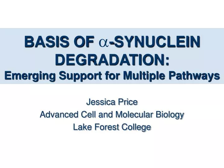
BASIS OF -SYNUCLEIN DEGRADATION:Emerging Support for Multiple Pathways Jessica Price Advanced Cell and Molecular Biology Lake Forest College
Road Map • Introduction to Parkinson’s Disease • -Synuclein Biology • Protein Degradation • Hypothesis • Results • Conclusions • Discussion & Future Research • Acknowledgments
Symptoms • Resting tremor • Muscular rigidity • Postural instability • Slowed movement • Also called bradykinesia http://www.michaeljfox.org/news/article.php?id=17&sec=2
Pathology • Death of dopaminergic neurons • Cytoplasmic inclusions of misfolded -synuclein called Lewy bodies http://www.med.harvard.edu/AANLIB/cases/case11/mr1/011.gif
-Synuclein Biology • 14 kDa protein • Abundant at presynaptic terminals • Precise function unknown • Mutations facilitate Lewy body aggregation in vitro http://medweb.bham.ac.uk/http/depts/clin_neuro/teaching/tutorials/parkinsons/lewy.jpg
Two forms of PD Sporadic (95%) Genetic (Familial) (5%) Mutations in: Environmental Factors • -Synuclein • UCHL-1 • Parkin • Park 3 • Pink-1 Misfolding ? Toxicity ? Aggregation (Lewy bodies) Cell Death
What happens to misfolded proteins? Extracellular and membrane proteins Proteins from the cytoplasm, nucleus, and ER http://www.nature.com/nrm/journal/v1/n2/slideshow/nrm1100_145a_F1.html
Ubiquitin-Proteasome System Colin Gordon, www.hgu.mrc.ac.uk/Research/Gordon.
Lysosome System All pathways involve vesicle mediated transport! Lysosome Figure 15-35. Biology 6th Edition, Campbell and Reece
Evidence for the Ub-proteasome -Synuclein is characterized as a cytoplasmic protein -Synuclein is degraded by the ub-proteasome pathway (Bennet et al., 1999; Holtz and O’Malley, 2003) Mutations associated with PD inhibit elements of the ub-proteasome pathway, Parkin, PARK 3, & ubiquitin C-terminal hydrolase L1 (Ceichanover and Brundin 2003; McNaught et al., 2002)
However, • Pharmacological studies have indicated that proteasome inhibitors do not alter cellular levels of α-synuclein (Rideout and Stefanis, 2002; Biasini et al., 2004) • -Synuclein has been shown to be translocated to lysosomes for degradation (Cuervo et al., 2004) • Wild type -Synuclein localizes to the cell membrane in yeast
The Lysosome: An alternate pathway? Willingham, et. al. 2003 identified 86 genes that increase -synuclein toxicity 32% were involved with vesicle mediated transport and lipid metabolism I chose to investigate Vps28
What does Vps28 do? • Component of the ESCRT-1 complex • ESCRT-1 recognizes Ub-cargo at the endosome and initiates transport of these cargos into vesicles that form MVBs
Hypothesis • The proteins composing the multivesicular body (MVB) sorting pathway play a key role in the transport of -synuclein to the lysosome for degradation.
Aim 1: • Verify -synuclein expression in cells lacking vps28 How? Western Analysis
Predictions Method: Western Analysis For all transformants a single band is expected at approximately 58 kDa, corresponding to the monomeric form of -synuclein tagged with GFP, when expression was induced by galactose.
-Synuclein is expressed in vps28 strains A30P/A53T, Gal pYES2, Gal GFP, Gal WT, Glu WT, Gal A30P, Gal A53T, Gal + - + - + - + - + - + - + - 1 2 3 4 5 6 7 8 9 10 11 12 13 14 + vps28 - vps28 58 kDa 1 2 3 4 5 6 7 8 9 10 11 12 13 14 36 kDa
Aim 2: • Assess the impact of the lack of vps28 on growth of -synuclein expressing cells How? Growth curve analysis & Dilution series spotting
Method: Grown Curve Analysis Method: Growth Curve Analysis Method: Growth Curve Analysis Glucose Evaluate OD over a period of 24h Galactose 24 h 24 h Prediction: Growth in all transformants lacking vps28 will be inhibited by the production of -synuclein, indicated by higher cell densities in transformants with vps28.
Method: Dilution Series Spotting 5X Less 5X Less 5X Less Prediction: Cells lacking vps28 will show inhibited growth when compared to the parent strain, with the mutant -synuclein transformants showing the most toxicity.
Spotting Assessment of Toxicity 5x dilutions 5x dilutions Vps28 + - + - + - WT α-synuclein pYES2 Plasmid GFP Non-Inducing Inducing
Spotting Assessment Cont. 5x dilutions 5x dilutions Vps28 + - + - + - A30P α-synuclein A53T α-synuclein A30P/A53T α-synuclein Non-Inducing Inducing
Aim 3: • Analyze the localization of -synuclein How? GFP Fluorescence Microscopy
Predictions Method: GFP Fluorescence Microscopy a-Synuclein will exhibit more cytosolic accumulation and aggregation in cells lacking vps28, with mutant a-Synucleins demonstrating greater levels of accumulation and aggregation.
Vps28 alters a-synuclein localization and increases aggregation Wild Type A30P A53T A30P/A53T + vps28 - vps28
Aim 4: • Assess the affect of vps28 absence on the persistence and stability of cells expressing a-synuclein How? Loss of Induction Assay
Method: Loss of Induction Assay Glucose Western Analysis Glucose Galactose 24 h 24 h 24 h Prediction: 0 2 4 6 8 10 12 14 16 18 Hours after Gal Shut-Off 58 kDa + Vps28 -Vps28 58 kDa
Vps28 does not appear to affect -synuclein stability over time Hours After Galactose Shut-Off 0 .5 1 2 4 6 9 12 18 24 58 kDa + Vps28 -Vps28 58 kDa
Hypothesis • The proteins composing the multivesicular body (MVB) sorting pathway play a key role in the transport of α-synuclein to the lysosome for degradation.
Conclusions • The absence of vps28 increases a-synuclein toxicity • Vps28 leads to a-synuclein accumulation in vivo • Vps28 presence does not discernibly alter a-synuclein clearance
Vps28 Absence increases a-Synuclein Toxicity • Increase in wild type a-synuclein toxicity previously been demonstrated in vps28in vivo by Willingham, et. al., 2003 confirmed • A30P, A53T, and A30P/A53T mutant a-synuclein toxicity was also modestly increased in the absence of vps28 • Absence variation in toxicity between wild type and mutant a-synucleins implies that the absence of vps28 is responsible for toxicity exclusively and not mutations in a-synuclein itself. • This explains the sporadic occurrence of PD in patients that do not have a-synuclein mutations, tying sporadic PD to the accumulation of a-synuclein due to dysfunctions in the vacuolar/lysosomal degradation pathway.
Vps28 leads to a-synuclein accumulation in vivo • Absence pf vps28 significantly alters the localization of all a-synuclein forms and increases the amount of a-synuclein cytoplasmic inclusion • Presence of cytoplasmic inclusions of all forms of a-synuclein in vps28 cells implies that the absence of vps28 leads to the accumulation of a-synuclein within the cell, a key aspect of PD. • The affect of vps28 on a-synuclein behavior points to the importance of the MVB pathway and the lysosome in a-synuclein degradation.
Vps28 presence does not discernibly alter a-synuclein clearance • Wild type a-synuclein persisted in both parent strain and vps28 cells • a-synuclein may be present in SDS-soluble aggregates which broke down to monomers • Lack of vps28 may not be enough to increase a-synuclein stability by a discernable amount • Impact on wild type a-synuclein stability may not be dramatic enough to capture in this assay
Discussion • The Ub-Proteasome System • The Lysosome System • A New Model
The Ub-Proteasome System:The Established Pathway • Ub-proteasome pathway degrades misfolded a-synuclein (Bennet et al., 1999; Holtz and O’Malley, 2003) • Dysfunction of this pathway linked to a-synuclein accumulation and aggregation (Sharma, 2004) • However, the function of the ubiquitin-proteasome in clearing a-synuclein from the cell has been brought into question, implicating an alternate method of a-synuclein degradation (Rideout and Stefanis, 2002; Biasini et al., 2004)
The Lysosome: The Emerging Pathway • a-Synuclein has also been shown to be targeted to and degraded by the vacuole/lysosome (Cuervo et al., 2004; Lee et al., 2004). • We demonstrated • Disruption of this pathway elevates the toxicity of all forms of a-synuclein • Disruption of this pathway increase a-synuclein accumulation and aggregation within cells • This indicates that disruption transport to the vacuole/lysosome for degradation has similar affects as the disruption of the ubiquitin-proteasome degradation pathway (Snyder et al., 2003, McNaught, et. al., 2003)
Vacuole/Lysosome Degradation MVB Pathway α-Synuclein Ubiquitin Established Pathway Misfolding Poly-Ub Mono-Ub Emerging Pathway
Two pathways work in conjunction to degrade a-synuclein Extracellular and membrane proteins Proteins from the cytoplasm, nucleus, and ER http://www.nature.com/nrm/journal/v1/n2/slideshow/nrm1100_145a_F1.html
Future Experiments • Quantify aggregation • Investigate other Vps proteins • DOA4 • Vps27 • Confirmation of the ubiquitination state of a-synuclein in vps28
Acknowledgments Dr. Shubhik DebBurman Isaac Holmes Nijee Sharma Katrina Brandis Sara Herrera Ruja Shrestha Lavinia Sintean Tasneem Saylawala Arun George Paul NIH NSF