Download
1 / 63
720 likes | 1.18k Views
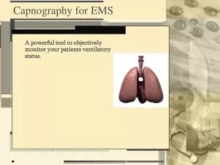
Applications of Capnography in EMS. Laura Kay MD Medical Director. Physiology. Oxygen -> lungs -> alveoli -> blood. Oxygen. breath. CO 2. muscles + organs. lungs. Oxygen. CO 2. cells. energy. blood. Oxygen + Glucose. CO 2. Physiology of Carbon Dioxide.

E N D
Applications of Capnography in EMS Laura Kay MD Medical Director
Physiology Oxygen -> lungs -> alveoli -> blood Oxygen breath CO2 muscles + organs lungs Oxygen CO2 cells energy blood Oxygen + Glucose CO2
Physiology of Carbon Dioxide ALL THREE ARE IMPORTANT! PERFUSION VENTILATION METABOLISM
CAPNOGRAPHY IS THE VITAL SIGN FOR VENTILATIONOXIMETRY IS THE VITAL SIGN FOR OXYGENTATION
ET Tube Verification(It can be harder than we think) RSI Inexperience Blood Facial Trauma Syringe/Bulb Trachlight Auscultation Infant/child I just know Combitube Seizures Experience Movement Short/fat neck Vomitus Mucus Recreational Drugs
Tube Vigilance………….“I know that tube was in!” • Moved to gurney • Moved to and from ambulance • CPR • Defibrillations • Moved to ED • Seizures • Agitated
Pearl Recent studies have shown that using a Cervical collar or head block dramatically decreases the incidence of tube displacement
Review of Ventilation/Respiration • Ventilation: Movement of gas in and out of the lungs • Diffusion: The passive two-way transfer of respiratory gases from the alveoli and plasma
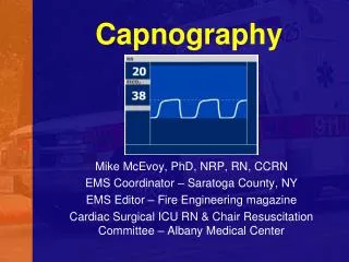
Normal Endtidal CO2 Normal 33-43 mmHg Waveform reflects how close numerical value is to actual end tidal volume Square - GOOD Hump - BAD
Expanding our Use of Capnography • Acute exacerbation of COPD • Asthma • Pneumonia • CHF • Respiratory Muscle Fatigue • Hypoventilation Syndromes • IS THE PATIENT RESPONDING TO THERAPY OR NOT?
Waveforms are a window into the patient • Square vs. Humped in appearance • Waveform shape compared to numeric value • Waveform shape, Numeric Value, Patient’s symptoms
Application to RSI • Cleft in the alveolar plateau indicates minimal spontaneous diaphragmatic movement • Partial recovery from neuromuscular blockade
Abnormal Waveforms • Gradual increase in EtCO2 • Rising body temperature • Hypoventilation • Increased metabolism
Abnormal Waveforms • Sustained low EtCO2 with a good plateau indicates either hyperventilation or a large physiological dead space ventilation, resulting in a widened a-ADCO2. • Pulmonary Emboli • Hypovolemia • Hyperventilation • COPD resulting in alveolar over-distension • Excessive level of PEEP
Abnormal Waveforms • Absent alveolar plateau indicates incomplete alveolar emptying or loss of endotracheal airway integrity. • Bronchospasm • Partial airway obstruction caused by secretions • Leak in the airway system • Partial disconnect from ventilator • Endotracheal tube in the hypopharynx
Trending • Trending of capnography provides a continuous view of the patient’s status- video versus snapshot • Early detection allows for early intervention • Trending is a simple tool that does not stress an already failing system
Hypoventilation Syndromes • Drug overdose • ETOH overdose • DKA • Post ictal • CVA • Head trauma • Neuromuscular
Factors influencing HypoventilationPatient at risk of Use of ETOH/Drugshypoventilation (?DX of OSA) Supplemental 02 in Oxygenation use slows desaturation does not = response ventilation Limited ability to Sedatives/injuriescommunicate impair cognitionStaff performing Patient has multiple several tasks injuries/other critical needs
Perfusion • Blood Flow (Perfusion) • Perfusion to the lungs • Skin color • Improving vital signs • HR • RR • BP • LOC
ETCO2 DURING CPR Onset of arrest ETCO2 decreases During CPR ETCO2 increases slightly ROSC ETCO2 markedly increases ROSC(cont’d) ETCO2 falls slightly Dependent on down time and preexisting conditions
Metabolism Pain Hyperthermia Malignant hyperthermia Shivering Circulatory System Increased cardiac output - with constant ventilation Respiratory System Respiratory insufficiency Respiratory depression Obstructive lung disease Equipment Defective exhalation valve Causes of an Elevated ETCO2
Metabolism Overdose / sedation Hypothermia Circulatory System Cardiac arrest Embolism Sudden hypovolemia or hypotension Respiratory System Alveolar hyperventilation Bronchospasm Mucus plugging Equipment Leak in airway system Partial airway obstruction ETT in hypopharynx Causes of a Decreased EtCO2
Case Study #1 • 32 yo 90 kg female presents in acute respiratory distress • Cyanotic, short word sentencing • Respiratory rate is shallow and labored at 24 • Expiratory phase is prolonged due to gas trapping • Heart Rate of 140, strong and bounding at the radial • BP 170/88
Patient is clutching her Albuterol inhaler (self-administered 15 puffs prior to yourarrival)ETC02 value was beginning to rise EtC02: 52 mmHg
Treatment • BLS assisted ventilations via Bag Valve Mask with 100% O2 and albuterol • Epinephrine 1:1000 SQ admin • Paramedic prepares for nasal intubation
Treatment • As the nasal tube is being advanced • The waveform below is obtained
What now? • Options?
Tube Confirmation • Cords visualized during intubation • Patient is successfully intubated orally • Auscultation reveals no discernible breath sounds anterior chest wall • Abdominal auscultation is equally silent • Minimal chest rise • Positive misting and condensation in the tube • Bulb aspirated syringe flows free-air
What is the next step? • Auscultation doesn't really help • SpO2 is not reading (before or after) • Remains difficult to ventilate. • What other tool can you use to confirm the placement of the tube? • What else could be going on with this patient? • Are we 100% sure we are in the trachea?
YES!!! We have an ETCO2 EtC02=22 mm Hg
Case # 2 • You respond to a 42you male involved in a truck vs motorcycle MVA • The patient is rapidly extricated, immobilized with a head block, on a long spine board • En route , the assessment is as follows:
Assessment • Head: large, bleeding scalp laceration • R pupil 4mm, sluggish, L pupil 8mm NR • + broken teeth with bleeding in airway • Chest atraumatic, clear breath sounds B and equal Abdomen soft, nondistended GCS 5-7
Treatment • The patient is orally intubated and the tube secured • The patient has a generalized tonic clonic seizure which lasts for approx. 2 minutes
The waveform is as follows • Corresponding waveform: C02 before the patient seized After the patient seized
Where is the tube???? • Provider checked the number at the lip • It had not changed • Listened to Breath Sounds • They were less audible • The EtC02 was 25, then dropped to 0 • Normal EtC02 is between 33 to 43 mm Hg
The Endotracheal tube was removed and the patient was reintubated • Corresponding waveform • CO2 now back up to 25mmHg
Case Study #3 • A 12 year old boy presented in acute respiratory failure with copious secretions and was successfully orally intubated and placed on a ventilator. • The patient remained obtunded, cyanotic and had little airway movement on auscultation.
Presentation • Unconscious & unresponsive • Respiration's unassisted remain agonal • Heart rate of 136 strong and regular at the radial • Blood Pressure 138/56 • ETCO2 32 mm HG What does this waveform show?
Answer: A slow upstroke and incomplete emptying- What could you do therapeutically?
The patient was suctioned and given a bronchodilator treatment What does this waveform show?
The waveform shows less obstruction and more of an alveolar plateau = improvedair movement Answer:
Case Study # 4 70 year old 60 kg female, status post cardiac arrest resuscitation by EMS She was resuscitated in the field and now being transported to hospital. Enroute Pt. is unconscious & unresponsive (orally intubated and on a vent) Vital Signs No spontaneous respirations BP 76/palpated Heart rate is palpable only at the carotid at 136 weak and irregular
Cardiac monitor reveals Sinus Arrhythmia with multi-focal PVC's and couplets • As you are debating Dopamine you notice a change... EtC02 drops from 30 to 18 mm Hg
Patient’s perfusion decreases more………….CO2 dropsfurther CPR is Initiated Now unable to palpate a carotid pulse
CPR was continued and palpable pulses with compressions were present. The improved waveform: