Download
1 / 43
430 likes | 443 Views
Discover the key features and functions of the skeletal system, including bone classifications, bone anatomy, bone tissue, and bone growth. Learn about common bone disorders such as osteoporosis and how to prevent them.

E N D
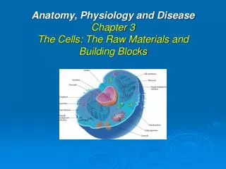
Health Sciences & OccupationsAnatomy, Physiology and Disease Chapter 6The Skeletal System
Introduction • Skeletal system - provides support and allows us to move • Bones (osseous tissue) - protects soft body parts - produces blood cells - acts as storage unit for minerals & fat • 206 bones in adult skeleton - along with cartilage, ligaments, and joints
Bones • Word ‘bone’ comes from Greek meaning “dried up body.” • Composed of non-living minerals such as calcium and phosphorous, bones are very much alive, constantly building and repairing themselves.
Bone Classifications • Long Bones: found in arms and legs • Short Bones: equal in width and length & found mostly in wrists and ankles • Flat Bones: thinner, flat or curved; can be plate-like & would include skull, ribs, and sternum (breast bone) • Irregular Bones: odd in shape, and include hip bone and vertebrae
Periosteum: Tough and fibrous connective tissue covering bone. Contains lymph vessels & nerves & blood vessels which transport blood & nutrients to nurture bone cells. Acts as anchor point for ligaments and tendons. Bone Anatomy
Bone Anatomy Con’t Epiphysis & Diaphysis • Epiphysis: formed by increase in size of both ends of long bone. • Diaphysis: region between two epiphyses • Medullary Cavity: hollow area acts as storage area for bone marrow. • Bone Marrow: • Yellow marrow: - has high fat content - can convert to red marrow in an emergency. • Red marrow: - produces red blood cells
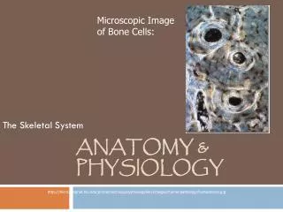
Bone Tissue • Dense, hard tissue that composes shafts of long bones. • Forms microscopic, cylindrical shaped units called osteons, or Haversian systems
Bone Tissue Con’t • Osteocytes: mature bone cells forming concentric circles around blood vessels • Area around osteocyte is filled with proteinfibers, calcium, and other minerals • Osteons run parallel to each other with blood vessels literally connecting with them to ensure sufficient oxygen and nutrients for bone cell
Bone Tissue Con’t Spongy (cancellous) bone • Arranged in bars and plates called trabeculae • Irregular holes between trabeculae make bone lighter in weight and provide space for red bone marrow, which produces red blood cells • Holesgive bone spongy appearance
Surface Structure of Bones Bone is not perfectly smooth • Projections act as points of attachment for muscles, ligaments, or tendons • Grooves and depressions act as pathways for nerves and blood vessels
Bone Growth and Repair Epiphyseal plate (growth plate) • After birth epiphysis on long bones continues to grow. • Plate is thin band of cartilage formed between primary and secondary ossification centers. • Plate exists as long as bones need to lengthen and widen; controlled by hormones, plate will eventually ossify and stop growth process.
Osteoporosis • As we age breakdown of bone becomes greater than formation of new bone (causing bone mass to gradually decrease). • Bones become lighter & weaker • holes in spongy bone becoming more prominent • weakened bones more prone to breakage • decreasing bone density called osteoporosis
Rx for Osteoporosis • Increase calcium: forms matrix of bone • Increase fluoride, & vitamin D: helps body absorb ingested calcium from digestive tract. • Stop smoking and decreasing caffeine consumption: both aid in calcium depletion. • Do weight-bearing exercise • Take medications to increase bone mass such as alendronate/Fosamax.
Cartilage • Connective tissue that can withstand fair amount of flexing, tension, and pressure: Nose & Ears. • Flexible connection between bones, as between ribs and sternum, allowing chest flexion during deep breathing & CPR.
Cartilage Cont • Acts as cushion between bones; “articular” cartilage located on ends of bones and acts as shock absorber/anti-grinding. • Bursa: small sacs secrete lubricant called synovial fluid. • Osteoarthritis: Inflammation of joints
Joints and Ligaments • Joint or Articulation: When two or more bones join together. • Ligaments: Tough, whitish bands that connect bone to bone. • Tendons: cord-like structures that attach muscle to bone.
Types of Synovial Joints • Pivot Joint: turnstile movement in neck and forearm • Ball and socket joint: hip and shoulder mvt rotation • Hinge joint: allow opening and closing movement in knees and elbows. • Gliding joint: wrists and ankles; provides sliding back and forth movement
Movement Classifications • Flexion: bending a joint • Extension: straightening a joint • Plantar flexion: pointing toes down • Dorsiflexion: bending foot up toward body • Abduction: moving away from body’s midline • Adduction: moving toward midline of body
Movement Classifications con’t • Inversion: turning foot medial • Eversion: turning foot lateral • Supination: turning hand palm up • Pronation: turning hand palm down • Circumduction: circular arm movement of a pitcher
Common Skeletal Disorders Osteoarthritis • Etiology: joint cartilage “wears out” • S/S: painful inflammation • Dx: visual exam; X-ray • Rx: rest, analgesics, anti-inflammatory meds, steroid injections (into affected joint), surgical intervention joint replacement.
Common Skeletal Disorders cont Rheumatoid arthritis: • Etiology: autoimmune disease that attacks connective tissue; especially joints. • S/S: stiffness, swelling and pain joints; inflammation of synovial membrane; pronounced joint deformities. • Dx: visual exam; X-ray; antibody screening (rheumatoid factor). • Rx: aspirin; NSAID; corticosteroid meds; methotrexate; rest & range of motion exercises; surgical intervention (in extreme cases)
Common Skeletal Disorders cont Septic arthritis • Etiology: infection inside joint, usually preceded by penetrating joint wound, or pathogen carried in blood. • S/S: pain & edema; filling of joint with inflammatory exudates; destruction of joint & replacement with fibrous tissue & eventually bone. • Dx: visual exam; X-ray; fluid culture for infection • Rx: antibiotics; prevent septic arthritis with good “aseptic techniques.”
Common Skeletal Disorders cont Bursitis: • Inflammation of a bursa • Etiology: repetitive movement; strain; congenital defect; rheumatic diseases • S/S: pain on movement; limited range of motion; inflammation and swelling at affected site • Dx: visual exam; X-ray • Rx: rest; moist heat/cold therapy; analgesics; NSAID; corticosteroid injection at affected site; draining (of fluid).
Common Skeletal Disorders cont Cruciate ligament tears: • Tear in one or more of ligaments of knee • Etiology: trauma induced when leg is twisted, planted (weight bearing) leg receives anterior or posterior blow. • S/S: pain in knee; instability of knee; limited mobility • Dx: physical exam (especially joint stability tests); radiologic exam (especially MRI). • Rx: NSAID, rest, immobilization, surgical repair
Common Skeletal Disorders cont Gout: • Etiology: metabolic disease where uric acid levels become too high; causes uric acid crystals to deposit in joints • S/S: pain in affected joint with inflammation & palpable heat tenderness with edema. • Dx: visual examination; blood testing for excessive uric acid. • Rx: low fat & low protein diet, rest & immobilization, Meds: anti-inflammatory medications & allopurinol. Monitor blood uric acid levels.
Common Skeletal Disorders cont Kyphosis: • Etiology: osteoporosis • S/S: exaggerated curve of upper back (“humpback”); may lead to backache, dyspnea/pulmonary insufficiency • Dx: visual exam, X-ray • Rx: depends on age and severity; may include: exercise; bracing; surgery; electrical stimulation; weight control
Common Skeletal Disorders cont Osteomalacia/Rickets: softening of bone • Etiology: decreased mineralization of bone due to insufficient vitamin D, lack of sufficient sunlight, malabsorption conditions. • S/S: bone pain & deformity; loss of height • Dx: visual examination; bone scan • Rx: correct nutritional deficiency: increase vitamin D
Common Skeletal Disorders cont Plantar fasciitis: (runner’s heel) • Etiology: repetitive impact on heel, resulting in inflammation of connective tissue on plantar surface of foot • Predisposing Factors: high arches, flat feet, shoes with poor support, increased body weight, & sudden increase in activity • S/S: intermittent pain • Dx: X-ray • Rx: rest, ice; NSAID; padding heel; possible surgery
Common Skeletal Disorders cont Tendonitis: inflammation of tendon • Etiology: repetitive movement • S/S: pain on movement with inflammation of involved area • Dx: visual exam; x-ray • Rx: rest; application of moist heat/cold; NSAID; injection of affected site with corticosteroids.
Common Skeletal Disorders cont Osteomyelitis: • Etiology: infection in bone, often preceded from wound in skin; staphylococcus aureus a common pathogen. • S/S: sudden pain, swelling, heat, & tenderness of affected site; high fever & chills, nausea & malaise. • Dx: visual exam, culture wound for pathogen • Rx: antibiotics; surgical debridement
Types of Bone Fractures • Simple: (closed): break without puncture to skin • Compound: (open): bone has been pushed through skin • Hairline: does not completely break or displace bone • Spiral: caused by severe twisting of bone • Greenstick: incomplete breaks, more common in children • Comminuted: when bone has been fragmented or splintered