Download
1 / 43
430 likes | 504 Views
Role of double IUI in cases with high PreHCG PSV. BY DR. C.B.NAGORI. MD., DGO . DR. SONAL., MD. Dr. Nagori’s Institute for Infertility and IVF “KEDAR”, Opp. Petrol pump, Nr. Parimal garden, Ellisbridge, Ahmedabad 380006. Introduction.

E N D
Role of double IUI in cases with high PreHCG PSV BY DR. C.B.NAGORI.MD., DGO. DR. SONAL., MD. Dr. Nagori’s Institute for Infertility and IVF “KEDAR”, Opp. Petrol pump, Nr. Parimal garden, Ellisbridge, Ahmedabad 380006
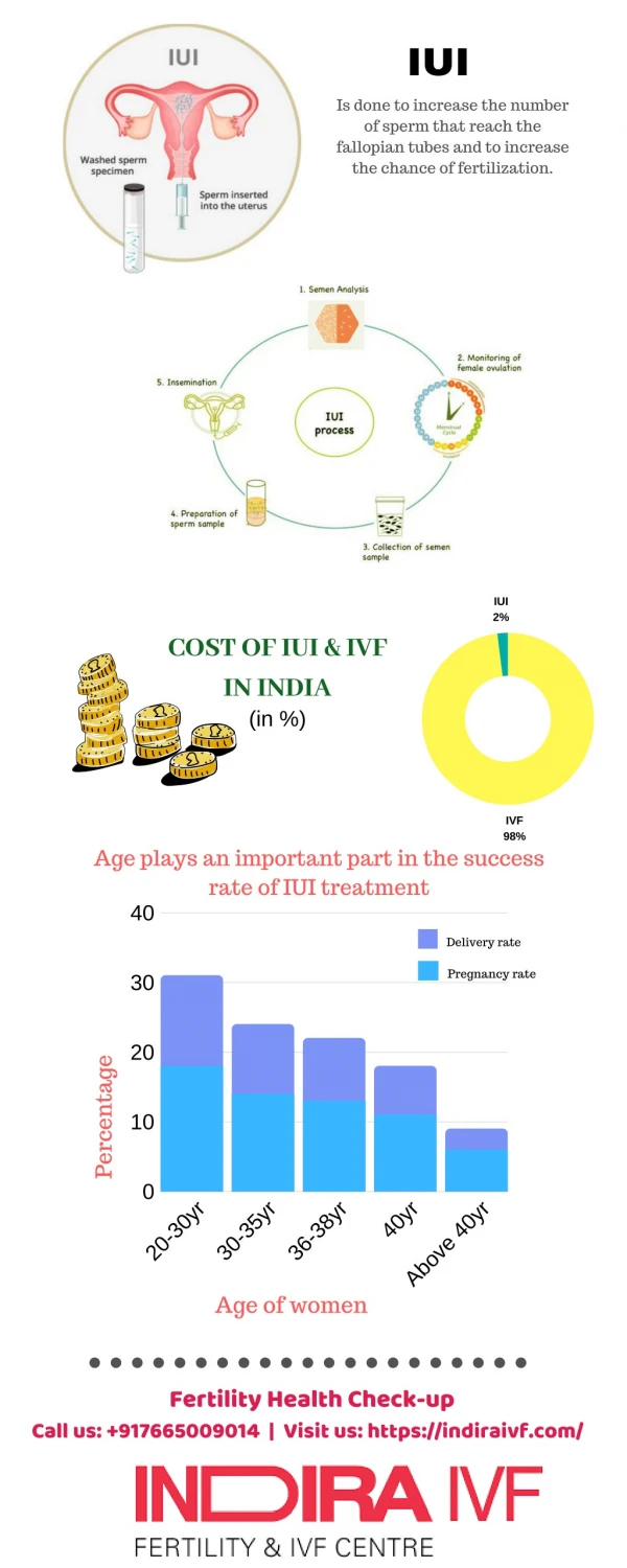
Introduction As has been studied by Prof. Kurjak and many other authors, the optimum perifollicular PSV, on the day of HCG has been confirmed as > 10cms/sec.
Vascular changes at the time of impending ovulation include increased vascularity of the inner wall of the follicle and a coincident surge in blood velocity just prior to erruption. Bourne et al, Intrafollicular blood flow during human ovulation, Ultrasound ObstetGynecol 1991; 1:53
A marked increase in the peak systolic velocity around the follicle, in the presence of a relatively constant PI, could be a sign of follicle maturity and impending ovulation. Tan SL, et al, Blood flow changes in the ovarian and uterine arteries during the normal menstrual cycle Am J ObstetGynecol 1996;175:625-31
Another study also says that the perifollicular PSV of 42cms/sec is reached about an hour before the spontaneous rupture of the follicle.
This means that if the follicle is said to be functionally mature when PSV is 10cms/sec, that is the time when the LH surge starts and under the effect of that LH, the perifollicular PSV keeps on rising constantly.
45cms/sec 10cms/sec
That means if higher the PSV, the follicle is closer to rupture.
Keeping this in mind, a study was done on patients undergoing IUI for their treatment of infertility.
Aim The aim of the study was to find out if double IUI can increase the pregnancy rate in patients who have a pre HCG perifollicular PSV > 15cms/sec.
Material and method 300 IUI cycles were included in the study. Patients were stimulated with clomiphene citrate, r FSH and letrazole + rFSH.
Monitoring was done with B mode as well as colourDoppler ultrasound. • All patients were denied intercourse when at least one follicle was more then 14mm in diameter.
Inclusion criteria • Patients with unexplained and dysovulatory infertility • Age group 22 – 40 years • Primary or secondary infertility of more than 3 years.
Exclusion criteria • Post wash count < 7 million / ml • Endometriosis grade 2 and 3 • Age > 40 years • More than two mature follicles
Method • Patients on stimulation were scanned by transvaginal route with 6-12MHz probe, on Voluson E8, Expert ultrasound machine (GE). • Patients were first scanned on 3rd day to assess the follicular size in each ovary and to assess the uterus for any abnormality. • After stimulation was started they were called on 7th day for reassessment and then intermittently till follicular size of 18mm is achieved.
Follicle is measured as single vertical diameter when it is seen as a circle. • If follicle appears oval or polygonal, three diameters are taken AP, transverse and longitudinal, in two sections perpendicular to each other and the mean of the three is taken as diameter. • When desired follicular diameter is achieved, the endometrium is evaluated.
Endometrial evaluation • Endometrial thickness of minimum 8 mm is considered as optimum. • It is measured as the broadest part of the endometrium, in the most longitudinal section of the uterus. • Endometrium is always measured from outer to outer margins of the echogenic margins of the endometrium. • The endometrium should be multilayered.
6mm, grade A/B Zone 3,4, RI < 0.6, PI 2 Vascular area> 5mm2 RI < 0.9, PI < 3 Uterine art Zaidi J et al, Endometrial thickness, morphology, vascular penetration and velocimetry in predicting implantation in an IVF program. Ultrasound ObstetGynecol 1995;6:191-8 Kupesic S, Kurjak A, et al, Luteal phase defect:comparison between dopplervelocimetry, histological and hormonal markers. Ultrasound ObstetGynecol 1997;9:105-12. Steer CV et al, Vaginal colourdoppler assessment on the day of ET accurately predicts patients in an IVF programme with suboptimal uterine perfusion who fail to become pregnant. Ultrasound ObstetGynecol 1991;1(Suppl):79-82 Volume > 3cc
When follicle and endometrium both met the required criteria on B mode ultrasound, colourdopplerassesment was done for follicle and endometrium. • For endometrium, when branches of spiral artery reached at least zone 3(hypoechoic area in between echogenic lines) and there were more than ten vessels reaching this zone, the endometrium was considered mature for implantation.
Follicular assessment: CD • Good follicle on colourdoppler was expected to have blood vessels covering more than 3/4th of its circumference and these blood vessels should have RI<0.48 and PSV> 10cms/sec. • Vessels, that obliterated the visualization of follicular walls are perifollicular vessels. • When these parameters are reached, injection of hCG is planned and IUI is done usually after 34-36 hours, routinely. • But in this study only those patients were included in whom the perifollicular PSV > 15 cms/sec, RI < 0.48.
RI < 0.5, PSV 10cms/s 16mm 3/4th vascularity Nargund G et al, Ultrasound derived indices of follicular blood flow before hCG administration and the prediction of oocyte recovery and preimplantation embryo quality. Human reprod 196; 11:2512-17 Nargund G et al, Associations between ultrasound indices of follicular blood flow, oocyte recovery and preimplantation embryo quality. Hum Reprod 1996; 11: 10-13 Bhal PS et al, Is follicular vascularity an index of pregnancy potential among women undergoing assisted reproduction treatment cycles? Hum Reprod 1997; 12:72
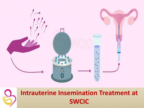
Method • hCG 10,000 was given to all patients for ovulation trigger. • Of all these patients, single IUI was done at 34-36 hours in half the patients and in half the patients, apart from the 34-36 hours IUI, an additional IUI was done at 12-14 hours. • Patient selection was at random for 50% each.
Method • HCG 10,000 was given for ovulation trigger when all these parameters were satisfied. • For half the patients with perifollicular PSV > 15 cms/sec, single IUI and for half double IUI was done randomly. • They were grouped into a PSV of 15 – 20, 20 - 25 and > 25 cms/sec.
Method • The results of the cycles were studied. • Conception was considered as a desired result and nonconception, an undesired result.
Observation Cycles total +ve -ve • Clomiphene citrate : 125 37 88 • rFSH : 125 54 71 • Letrazole + rFSH : 50 19 31
Conclusion • With any stimulation protocol when perifollicular PSV on the day of hCGis > 25cms/sec, double IUI must be done.
Conclusion • With rFSH and combination protocols with the PSV values > 20 cms/sec also double IUI must be preferred.
Conclusion • With PSV < 20 for any protocol, single and double IUI show no significant change in pregnancy rates.
THANK YOU DR. NAGORI’S INSTITUTE FOR INFERTILITY AND IVF, ‘KEDAR, NR.PARIMAL GARDEN, ELLISBRIDGE, AHMEDABAD. 380006