Download
1 / 1
10 likes | 112 Views
Discovery of 26 Panel Protein Biomarkers from Coupled Proteomics and Systems Biology Methods. Fan Zhang 1, 6 , Harikrishna Nakshatri 2,3,4 , Mu Wang 3,5,6 and Jake Y. Chen 1,6*.

E N D
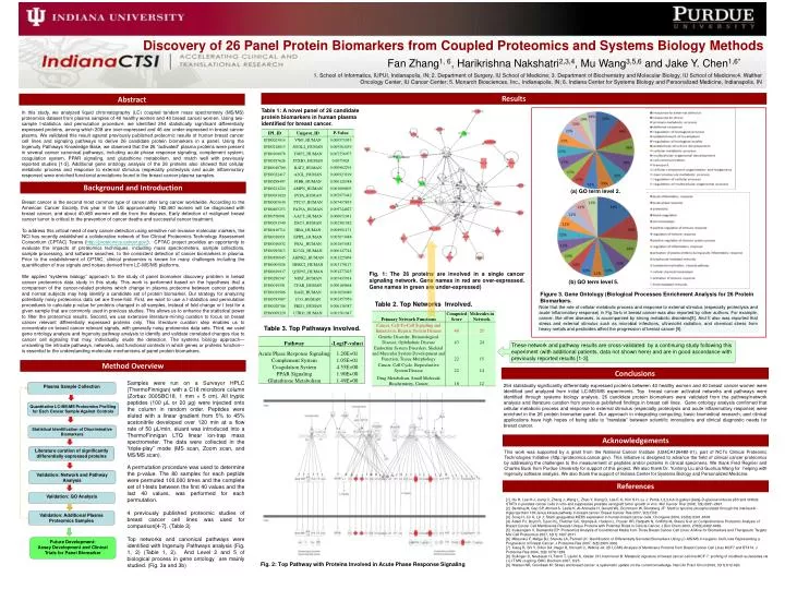
Discovery of 26 Panel Protein Biomarkers from Coupled Proteomics and Systems Biology Methods Fan Zhang1, 6, Harikrishna Nakshatri2,3,4, Mu Wang3,5,6 and Jake Y. Chen1,6* 1. School of Informatics, IUPUI, Indianapolis, IN; 2. Department of Surgery, IU School of Medicine; 3. Department of Biochemistry and Molecular Biology, IU School of Medicine;4. Walther Oncology Center, IU Cancer Center; 5. Monarch Biosciences, Inc., Indianapolis, IN; 6. Indiana Center for Systems Biology and Personalized Medicine, Indianapolis, IN Results Abstract Table 1: A novel panel of 26 candidate protein biomarkers in human plasma identified for breast cancer. In this study, we analyzed liquid chromatography (LC) coupled tandem mass spectrometry (MS/MS) proteomics dataset from plasma samples of 40 healthy women and 40 breast cancer women. Using two-sample t-statistics and permutation procedure, we identified 254 statistically significant differentially expressed proteins, among which 208 are over-expressed and 46 are under-expressed in breast cancer plasma. We validated this result against previously published proteomic results of human breast cancer cell lines and signaling pathways to derive 26 candidate protein biomarkers in a panel. Using the Ingenuity Pathways Knowledge Base, we observed that the 26 “activated” plasma proteins were present in several cancer canonical pathways, including acute phase response signaling, complement system, coagulation system, PPAR signaling, and glutathione metabolism, and match well with previously reported studies [1-3]. Additional gene ontology analysis of the 26 proteins also showed that cellular metabolic process and response to external stimulus (especially proteolysis and acute inflammatory response) were enriched functional annotations found in the breast cancer plasma samples. Background and Introduction (a) GO term level 2. Breast cancer is the second most common type of cancer after lung cancer worldwide. According to the American Cancer Society, this year in the US approximately 182,460 women will be diagnosed with breast cancer, and about 40,480 women will die from the disease. Early detection of malignant breast cancer tumor is critical to the prevention of cancer deaths and successful cancer treatment. To address this critical need of early cancer detection using sensitive non-invasive molecular markers, the NCI has recently established a collaborative network of five Clinical Proteomics Technology Assessment Consortium (CPTAC) Teams (http://proteomics.cancer.gov/). CPTAC project provides an opportunity to evaluate the impacts of proteomics techniques, including mass spectrometers, sample collections, sample processing, and software searches, to the consistent detection of cancer biomarkers in plasma. Prior to the establishment of CPTAC, clinical proteomics is known for many challenges including the quantification of true signals and noises derived from LC-MS/MS platforms. We applied “systems biology” approach to the study of panel biomarker discovery problem in breast cancer proteomics data study in this study. This work is performed based on the hypothesis that a comparison of the cancer-related proteins which change in plasma proteome between cancer patients and normal subjects may help identify a candidate set of protein biomarker. Our strategy for analyzing potentially noisy proteomics data set are three-fold. First, we want to use a t-statistics and permutation procedures to calculate p-value for proteins changed in all samples, instead of fold change or t test for a given sample that are commonly used in previous studies. This allows us to enhance the statistical power to filter the proteomics results. Second, we use extensive literature-mining curation to focus on breast cancer relevant differentially expressed proteins only. This literature curation step enables us to concentrate on breast cancer relevant signals, with generally noisy proteomics data sets. Third, we used gene ontology analysis and Ingenuity pathway analysis to identify and validate correlated changes due to cancer cell signaling that may, individually, elude the detection. The systems biology approach—unraveling the intricate pathways, networks, and functional contexts in which genes or proteins function—is essential to the understanding molecular mechanisms of panel protein biomarkers. Fig. 1: The 26 proteins are involved in a single cancer signaling network. Gene names in red are over-expressed. Gene names in green are under-expressed) (b) GO term level 5. Figure 3. Gene Ontology (Biological Processes Enrichment Analysis for 26 Protein Biomarkers. Table 2. Top Networks Involved. Note that the role of cellular metabolic process and response to external stimulus (especially proteolysis and acute inflammatory response) in Fig 3a-b in breast cancer was also reported by other authors. For example, cancer, like other diseases, is accompanied by strong metabolic disorders[8]. And It also was reported that stress and external stimulus such as microbial infections, ultraviolet radiation, and chemical stress from heavy metals and pesticides affect the progression of breast cancer [9]. Table 3. Top Pathways Involved. These network and pathway results are cross-validated by a continuing study following this experiment (with additional patients, data not shown here) and are in good accordance with previously reported results [1-3]. Method Overview Conclusions Samples were run on a Surveyor HPLC (ThermoFinnigan) with a C18 microbore column (Zorbax 300SBC18, 1 mm × 5 cm). All tryptic peptides (100 μL or 20 μg) were injected onto the column in random order. Peptides were eluted with a linear gradient from 5% to 45% acetonitrile developed over 120 min at a flow rate of 50 μL/min, eluant was introduced into a ThermoFinnigan LTQ linear ion-trap mass spectrometer. The data were collected in the “triple-play” mode (MS scan, Zoom scan, and MS/MS scan). A permutation procedure was used to determine the p-value. The 80 samples for each peptide were permuted 100,000 times and the complete set of t-tests between the first 40 values and the last 40 values, was performed for each permutation. 4 previously published proteomic studies of breast cancer cell lines was used for comparison[4-7]. (Table 3) Top networks and canonical pathways were identified with Ingenuity Pathways analysis (Fig. 1, 2) (Table 1, 2). And Level 2 and 5 of biological process in gene ontology are mainly studied. (Fig. 3a and 3b) 254 statistically significantly differentially expressed proteins between 40 healthy women and 40 breast cancer women were identified and analyzed from initial LC-MS/MS experiments. Top breast cancer activated networks and pathways were identified through systems biology analysis. 26 candidate protein biomarkers were validated from the pathway/network analysis and literature curation from previous published findings in breast cell lines. Gene ontology analysis confirmed that cellular metabolic process and response to external stimulus (especially proteolysis and acute inflammatory response) were enriched in the 26 protein biomarker panel. Our approach in integrating computing, basic biomedical research, and clinical applications have high hopes of being able to “translate” between scientific innovations and clinical diagnostic needs for breast cancer. Plasma Sample Collection Quantitative LC-MS/MS Proteomics Profiling for Each Cancer Sample Against Controls Statistical Identification of Discriminative Biomarkers Acknowledgements Literature curation of significantly differentially expressed proteins This work was supported by a grant from the National Cancer Institute (U24CA126480-01), part of NCI‟s Clinical Proteomic Technologies Initiative (http://proteomics.cancer.gov). This initiative is designed to advance the field of clinical cancer proteomics by addressing the challenges to the measurement of peptides and/or proteins in clinical specimens. We thank Fred Regnier and Charles Buck from Purdue University for support of this project. We also thank Dr. Yunlong Liu and Guohua Wang for helping with Ingenuity software analysis. We also thank the support of Indiana Center for Systems Biology and Personalized Medicine. Validation: Network and Pathway Analysis References Validation: GO Analysis [1] Hu H, Lee H-J, Jiang C, Zhang J, Wang L, Zhao Y, Xiang Q, Lee E-O, Kim S-H, Lu J: Penta-1,2,3,4,6-O-galloyl-{beta}-D-glucose induces p53 and inhibits STAT3 in prostate cancer cells in vitro and suppresses prostate xenograft tumor growth in vivo. Mol Cancer Ther 2008, 7(9):2681-2691. [2] Berishaj M, Gao SP, Ahmed S, Leslie K, Al-Ahmadie H, Gerald WL, Bornmann W, Bromberg JF: Stat3 is tyrosine-phosphorylated through the interleukin-6/glycoprotein 130/Janus kinase pathway in breast cancer. Breast Cancer Res 2007, 9(3):R32. [3] Song H, Jin X, Lin J: Stat3 upregulates MEK5 expression in human breast cancer cells. Oncogene 2004, 23(50):8301-8309. [4] Adam PJ, Boyd R, Tyson KL, Fletcher GC, Stamps A, Hudson L, Poyser HR, Redpath N, Griffiths M, Steers G et al: Comprehensive Proteomic Analysis of Breast Cancer Cell Membranes Reveals Unique Proteins with Potential Roles in Clinical Cancer. J Biol Chem 2003, 278(8):6482-6489. [5] Kulasingam V, Diamandis EP: Proteomics Analysis of Conditioned Media from Three Breast Cancer Cell Lines: A Mine for Biomarkers and Therapeutic Targets. Mol Cell Proteomics 2007, 6(11):1997-2011. [6] Mbeunkui F, Metge BJ, Shevde LA, Pannell LK: Identification of Differentially Secreted Biomarkers Using LC-MS/MS in Isogenic Cell Lines Representing a Progression of Breast Cancer. J Proteome Res 2007, 6(8):2993-3002. [7] Xiang R, Shi Y, Dillon DA, Negin B, Horvath C, Wilkins JA: 2D LC/MS Analysis of Membrane Proteins from Breast Cancer Cell Lines MCF7 and BT474. J Proteome Res 2004, 3(6):1278-1283. [8] Bullinger D, Neubauer H, Fehm T, Laufer S, Gleiter CH, Kammerer B: Metabolic signature of breast cancer cell line MCF-7: profiling of modified nucleosides via LC-IT MS coupling. BMC Biochem 2007, 8:25. [9] Nielsen NR, Gronbaek M: Stress and breast cancer: a systematic update on the current knowledge. Nat Clin Pract Oncol 2006, 3(11):612-620. Validation: Additional Plasma Proteomics Samples Future Development: Assay Development and Clinical Trials for Panel Biomarker Fig. 2: Top Pathway with Proteins Involved in Acute Phase Response Signaling