Download
1 / 42

E N D
INFLAMMATION DR.AYSER HAMEED LEC.1
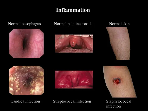
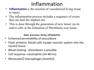
General Features Inflammation is defined as "the response of living vascularized tissues to harmful agents.”It consists principally of vascular changes associated with leukocytes infiltration and systemic reactions. Inflammation is a fundamental and common pathologic process seen in many disease states.
It is essentially a protective response, the aim of which is to get rid of the injurious agents (e.g., microbes, toxins) as well as its consequences (e.g. necrotic cells and tissues). Inflammation is concurrently tangled with another process (repair) that tries to mend the damaged tissues resulting from the battle between the offending agent and the host.
Without inflammation, infections would go uninhibited, wounds would never heal, and injured organs might remain permanently damaged. Sometimes, however, inflammation and its associated repair may be potentially harmful. For this reason, pharmacies flourish with anti-inflammatory drugs, which ideally control the harmful sequelae of inflammation yet do not interfere with its beneficial effects.
Many tissues and cells are involved in the inflammatory reaction, including plasma fluid proteins, circulating leukocytes, blood vessels, and cellular and extracellular constituents of connective tissues. The circulating leukocytes include neutrophils, monocytes, eosinophils, lymphocytes, basophils, in addition to platelets. The connective tissue cells are mast cells, fibroblasts, macrophages, and lymphocytes.
The extracellular matrix consists of structural proteins (collagen, elastin), adhesive glycoproteins (fibronectin, laminin), and proteoglycans. Inflammation is divided into acute or chronic. The latter includes also a specific form called granulomatous inflammation.
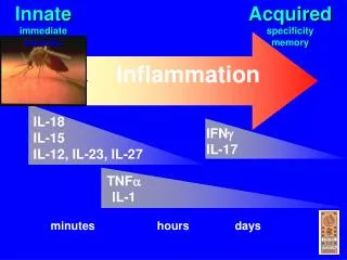
Acute inflammationis rapid in onset (seconds or minutes), of relatively short duration (minutes, hours, or at most a few days), characterized by the exudation of fluid and plasma proteins & the emigration of leukocytes, predominantly neutrophils. Chronic inflammationin contradistinction, is of insidious onset, of longer duration, and is associated histologically with the presence of lymphocytes, macrophages, plasma cells, proliferation of blood vessels and fibroblasts.
In both forms tissue necrosis of varying extent occurs. The vascular and cellular reactions of both acute and chronic inflammation are mediated by chemical substances (chemical mediators) that are derived from plasma proteins or cells. Such substances, acting singly, in combinations, or in sequence, amplify the inflammatory response and influence its evolution.
The five cardinal signs of inflammation Are rubor (redness), tumor (swelling), calor (heat), dolor (pain), and loss of function (functiolaesa). The first four signs are typically more prominent in acute inflammations than in chronic ones.
ACUTE INFLAMMATION Stimuli of acute inflammation Acute inflammatory reactions are triggered by a variety of stimuli that include:- 1. Infections: bacterial, viral, parasitic and microbial toxins. 2. Physical and chemical agents (trauma, thermal injuries, irradiation, toxins, strong acids, etc.). 3. Tissue necrosis (of any from or cause). 4. Foreign bodies (splinters, dirt, sutures). 5. Immune reactions (hypersensitivity and autoimmune reactions).
Exudationis the escape of fluid, proteins, and blood cells from the vascular system into the interstitial tissue. An exudate is an extravascular fluid that has a high protein concentration and a specific gravity above 1.020. It involves significant alteration in the normal permeability of small blood vessels in the area of injury. In contrast, a transudateis a fluid with low protein content (most of which is albumin) and a specific gravity of less than 1.012.
It is essentially an ultrafiltrate of blood plasma that results from osmotic or hydrostatic imbalance across the vessel wall without an increase in vascular permeability. Edema refers to an excess of fluid in the interstitial tissues or body cavities; the accumulated fluid can be either an exudate or a transudate. Pus (purulent exudate) is an inflammatory exudate rich in leukocytes (mostly neutrophils), the debris of dead cells and, in many cases, microbes (pyogenic bacteria).
Acute inflammation has three major components: A. Vasodilation associated with increased blood flow. B. Increased vascular permeability associated with decreased blood flow. C. Emigration and activation of leukocytes and phagocytosis.
The major local manifestations of acute inflammation, compared to normal • Vascular dilation and increased blood flow (causing erythema and warmth), • extravasation and deposition of plasma fluid and proteins (edema), and • leukocyte (mainly neutrophil) emigration and accumulation in the site of injury.
A.Vasodilation and increased blood flow: This is, sometimes, preceded by a transient constriction of arterioles, lasting a few seconds. Vasodilation first involves the arterioles, which leads to an increase in blood flow; this in turn leads to opening of new capillary beds in the area with subsequent dilation of capillaries & venules.
This process allows more blood to flow into the area, a process known as “active hyperemia” (hyper- = increased; -emia = blood). These changes explain the clinically noted heat and redness. Vasodilation is induced by the action of several mediators (such as histamine) on vascular smooth muscles. It is possible that autonomic nerve impulses may also play a role in relaxation of arteriolar smooth muscle leading to their dilation.
B.Increased Vascular Permeability and decreased blood flow: Increased vascular permeability leads to the escape of exudates into the extravascular tissue. This is driven by the increased hydrostatic pressure owing to increased blood flow through the dilated vessels and is perpetuated through the loss of proteins from the plasma that reduces the intravascular osmotic pressure and increases the osmotic pressure of the interstitial fluid. Several mechanisms have been proposed for the increased vascular permeability that includes:
1. Formation of endothelial gaps in venules due to endothelial cells contraction. This is the most common mechanism & is elicited by several mediators e.g. histamine, bradykinin, and leukotrienes. Binding of these mediators to receptors on endothelial cells leads to stimulation of contractile proteins (such as myosin). The result is contraction of the endothelial cells and separation of intercellular junctions that eventuate in intercellular gaps formation. 2. Junctional retraction caused bychemical mediators such as TNF and IL-1; these induce structural reorganization of the cytoskeleton of the cells.
3. Direct endothelial cell injury as by burns or infections. Because of endothelial damage and exposure of the subendothelialthrombogenic collagen, this type is frequently associated with platelets adhesion with subsequent thrombosis. 4. Leukocyte-dependant injury due to accumulation of leukocytes and their activation products (such as toxic oxygen radicals and proteolytic enzymes) during the inflammatory response. These lead to endothelial cell damage.
According to the above mechanisms, there are three basic patterns of increased permeability: 1. Immediate transient response lasting for 30 minutes or less, mediated mainly by the actions of histamine and leukotrienes on endothelium. 2. Delayed response starting at about 2 hours and lasting for about 8 hours, mediated principally by kinins, complement products. 3. Prolonged response that is most noticeable after direct endothelial injury, e.g. after burns.
The inflammatory exudate, in addition to leukocytes, is composed of plasma proteins; of these, two play a particularly important role: 1. Immunoglobulins; a group of antibodies that have the ability to react with certain antigens, making them vulnerable to the actions of neutrophils and macrophages (opsonization). 2. blood clotting proteins; a blood clot is composed of a meshwork of "fibrin"a protein end product of a complex interaction of plasma, tissues, and cell factors . The fibrin so produced prevents spread of the injurious agents.
Plasma leakage has an effect inside blood vessels as well. Blood cells become more closely packed (hemoconcentration) causing sluggish flow or even complete stasis. When blood flow is normal the "formed elements" are found in a cell-rich "axial core" being separated from the endothelial lining by a thin cell-free "plasmic zones." As blood flow slows down during inflammation, the axial core can no longer be maintained; this allows blood leukocytes to come in contact with the endothelial lining cells (margination).
C. Emigration and activation of leukocytes and phagocytosis. A critical function of inflammation is to deliver leukocytes to the sites of injury and to activate the leukocytes to defend the host. Leukocytes ingest offending agents, kill bacteria and other microbes, and get rid of necrotic tissues and foreign substances. However, these cells may induce tissue damage and prolong inflammation. The journey of leukocytes from the vessels lumen to the interstitial tissue is called extravasation. This can be divided into the following steps:
1. Binding of leukocytes to the endothelial cells. Normally, the vascular endothelium does not bind circulating cells or impede their passage. In inflammation, however, the endothelium becomes activated to permit binding of leukocytes to its surface. This is followed by. 2. Transmigration of leukocytes across the endothelium (diapedesis). 3. Migration of leukocytes within the interstitial tissues toward the focus of tissue injury.
Because blood flow slows down in inflammation, more white cells assume a peripheral position along the endothelial surface. This process is called margination. Subsequently, leukocytes tumble and roll over slowly along the endothelium and eventually come to rest through firm adhesions with the endothelial cells. In time, the endothelium becomes virtually lined by white cells, an appearance called pavementing. After firm adhesion, leukocytes insert pseudopods into the junctions between the endothelial cells, squeeze through interendothelial junctions, and eventually, traverse the basement membrane and escape into the extravascular space.
Neutrophils, monocytes, lymphocytes, eosinophils, and basophils, all use the same pathway to migrate from the blood into tissues. Leukocyte adhesion and transmigration are achieved by the binding of complementary adhesion molecules on the leukocyte and endothelial surfaces, a process regulated by chemical mediators. The adhesion receptors involved belong to several molecular families including selectins and integrins. The next step in the process is migration of the leukocytes through the endothelium, called transmigration or diapedesis.
Chemokines (chemoattractants) act on the adherent leukocytes and stimulate the cells to migrate toward the site of injury or infection. Certain adhesion molecules, present in the intercellular junction of endothelium, are involved in the migration of leukocytes. Leukocyte diapedesis, similar to increased vascular permeability, occurs predominantly in the venules. After traversing the endothelium, leukocytes eventually pierce the basement membrane, probably by secreting degrading enzymes such as collagenases & elastases.
Once leukocytes enter the extravascular connective tissue, they are able to adhere to the extracellular matrix by virtue of integrins and CD44. Thus, the leukocytes are retained at the site where they are needed. The type of emigrating leukocyte varies with the age of the inflammatory response and with the type of stimulus. In most forms of acute inflammation, neutrophils predominate in the inflammatory infiltrate during the first 6 to 24 hours, and then are replaced by monocytes in 24 to 48 hours.
After entering tissues, neutrophils are short-lived; they undergo apoptosis (self destruction) and disappear after 24 to 48 hours, whereas monocytes (by now called macrophages: macro- = large and phage = eater) survive longer and thus outlive neutrophils and become more apparent. There are, however, exceptions to this pattern of cellular exudation.
In certain infections—for example, those produced by Pseudomonas organisms—neutrophils predominate over 2 to 4 days; in viral infections, lymphocytes may be the first cells to arrive; in some hypersensitivity reactions and parasitic infestations, eosinophils may be the main cell type.
Chemotaxis After extravasation, leukocytes emigrate in tissues toward the site of injury; this is achieved by a process called chemotaxis. Chemotaxis is defined as locomotion oriented along a chemical gradient of chemoattractants.
All granulocytes, monocytes and, to a lesser extent, lymphocytes respond to chemoattractants (chemotactic stimuli) with varying rates of speed. Both exogenous and endogenous substances can act as chemoattractants. The former is exemplified by bacterial products.
Endogenous chemoattractants, however, include several chemical mediators: 1. Components of the complement system, particularly C5a. 2. Products of the lipoxygenase pathway, mainly leukotriene B4 (LTB4). 3. Cytokines (secreted from cells) e.g., IL-8.
All the chemoattractants mentioned above bind to specific receptors on the surface of leukocytes. Signals initiated from these receptors result in recruitment & activation of specific leukocytic proteins including tyrosine kinases.
These changes eventuate in polymerization of actin that results in increased amounts of this contractile protein at the leading edge of the cell. The leukocyte moves by extending filopodia that pull the back of the cell in the direction of extension, much as a car with front-wheel drive is pulled by the wheels in front.
Leukocyte Activation This refers to induction of a number of responses within leukocytes, which are mediated by:- • Microbes. • Products of necrotic cells. • Antigen-antibody complexes. • and cytokines. These mediators trigger several signaling pathways in leukocytes that result in an increase in cytoplasmic Ca++ and activation of enzymes.
The activation of leukocytes is reflected functionally as follows: 1. Production of arachidonic acid (AA) metabolites. 2. Secretion of lysosomal enzymes and other microbicidal substances. 3. Modulation of leukocyte adhesion molecules allowing firm adhesion to endothelium.
4. Activation of macrophages: through the release of IFN-ɤ (major macrophage-activating cytokine), which is secreted by natural killer (NK) cells. 5. Activation of phagocytosis through stimulation of opsonins-receptors. The process of coating a particle, such as a microbe, to make it vulnerable for phagocytosis is called opsonization; substances that do this are opsonins.
Phagocytosis Phagocytosis is one of the major functions of the accumulated neutrophils and macrophages at the inflammatory focus, being responsible for eliminating the injurious agents. Phagocytosis involves three distinct but interrelated steps: 1. Recognition and attachment of the particle to be ingested by the leukocyte. 2. Its engulfment, with subsequent formation of a phagocytic vacuole. 3. Killing and degradation of the ingested material.
Recognition and Attachment Although neutrophils and macrophages can engulf bacteria without attachment to specific receptors, typically the phagocytosis of microbes and dead cells is initiated by recognition of these particles by receptors expressed on the leukocyte surface.
The efficiency of phagocytosis is greatly enhanced when microbes are opsonized by specific proteins (opsonins) for which the phagocytes express high-affinity receptors. The major opsonins are IgG antibodies, the C3b breakdown product of complement, and certain plasma lectins.