Download

1 / 76
770 likes | 1.04k Views
LEUKAEMIA DIAGNOSIS. Leukaemia is a disease resulting from the neoplastic proliferation of haemopoietic or lymphoid cells. Leukaemias are broadly divided into: Acute leukaemias, which, if untreated, lead to death in weeks or months.

E N D
LEUKAEMIA DIAGNOSIS
Leukaemia is a disease resulting from the neoplastic proliferation of haemopoietic or lymphoid cells. Leukaemias are broadly divided into: • Acute leukaemias, which, if untreated, lead to death in weeks or months. • chronic leukaemias, which, if untreated, lead to death in months or years.
They are further divided into lymphoid, myeloid and biphenotypic leukaemias, the latter showing both lymphoid and myeloid differentiation. Acute leukaemias are characterized by a defect in maturation, leading to an imbalance between proliferation and maturation; since cells of the leukaemic clone continue to proliferate without maturing to end cells.
Diagnosis of leukaemia. The diagnosis of leukaemia and categorization required the following parameters. 1- Morphology. 2-Cytochimestry 3-Immunophenotyping. 4-Cytogenetic. 5- Molecular study.
Background Acute lymphoblastic leukemia (ALL) is the most common malignancy diagnosed in children, representing nearly one third of all pediatric cancers. The annual incidence rate for acute lymphoblastic leukemia is 30.9 cases per million population. The peak incidence occurs in children aged 2-5 years.
Pathophysiology In acute lymphoblastic leukemia, a lymphoid progenitor cell becomes genetically altered and subsequently undergoes dysregulated proliferation, survival, and clonal expansion. In most cases, the pathophysiology of transformed lymphoid cells reflects the altered expression of genes whose products contribute to the normal development of B cells and T cells
Clinical feature. Children with acute lymphoblastic leukemia (ALL) generally Present with signs and symptoms that reflect bone marrow infiltration and extramedullary disease. Because leukemic blasts replace the bone marrow, patients Present with signs of bone marrow failure, including anemia, thrombocytopenia, and neutropenia. Clinical manifestations include fatigue and pallor, petechiae and bleeding, and fever. In addition, leukemic spread may manifest as lymphadenopathy And hepatosplenomegaly. Other signs and symptoms of leukemia including weight loss, bone pain, and dyspnea.
The classification of ALL FAB classification. L1 ALL L2 ALL L3 ALL Cell size Mainly small Large, heterogeneous Large, homogeneous Nuclear chromatin Fairly homogeneous Heterogeneous Finely stippled, Nuclear shape Mainly regular Irregular; clefting Regular Nucleolus Not visible Usually visible Usually prominent Amount of cytoplasm Scanty Variable abundant Moderately abundant Cytoplasmic basophilia Slight to moderate Variable Strong Cytoplasmic vacuolation Variable Variable Often prominent
Clinical correlates of FAB categories of ALL Many cases of L3 ALL represent a distinct entity that requires specific management. However, the categorization of a case as L1 or L2 ALL is of little importance. The FAB L1 category includes more childhood cases with a relatively good prognosis. The incidence of ALL L1 falls with increasing age whereas the incidence of ALL L2 does not vary much with age. ALL L2 has generally been found to have a worse prognosis, although the difference is not major.
WHO proposed classification of acute lymphoblastic leukemia The recent WHO International panel on ALL recommends that the FAB classification be abandoned, since the morphological classification has no clinical or prognostic relevance. 1- Acute lymphoblastic leukemia/lymphoma Synonyms: Former Fab L1/L2 i. Precursor B acute lymphoblastic leukemia/lymphoma. Cytogenetic subtypes: • t(12;21)(p12,q22) TEL/AML-1 • t(1;19)(q23;p13) PBX/E2A • t(9;22)(q34;q11) ABL/BCR • T(V,11)(V;q23) V/MLL ii. Precursor T acute lymphoblastic leukemia/lymphoma 2- Burkett's leukemia/lymphoma Synonyms: Former FAB L3 3- Biphenotypic acute leukemia
Characterization of the Immunophenotyping is referred to as Immunophenotyping and is achieved by means of labeled antibodies that recognize specific epitopes of cellular antigens. In general, the most useful antibodies are monoclonal antibodies (McAb) produced by hybridoma technology but, for some antigens, polyclonal antibodies (PcAb) (antisera) are better. The technique employed for Immunophenotyping may be immunocytochemistry or, much more often, flow cytometry. Immunophenotyping is essential for the diagnosis of B- or T-lineage acute lymphoblastic leukaemia (ALL).
First panel B lymphoid CD19, CD22, CD79a, CD10 T lymphoid CD3, CD2, CD7 Second panel If B lineage cm, k, l, CD20, CD24 If T lineage CD1a, SmCD3, CD4, CD5, CD8, anti-TCR ab, anti-TCR gd
Cytogenetic study. With techniques now available, 70–90% of cases of ALL have a demonstrable cytogenetic abnormality. In ALL, chromosomal abnormalities correlate with other clinical and hematological factors of prognostic importance but they also have a considerable independent prognostic significance.
B-lineage ALL L1 high/ hyperdiploidy. L1 or L2/t(9;22)/BCR-ABL fusion L1 or L2/t(4;11)(q21;q23) L1 or L2/t(12;21)(p12;q22)/early precursor or common ALL L1 or L2/t(1;19)(q23;p13)/pre-B ALL
T-lineage ALL. L1 or L2/t(10;14)(q24;q11) Burkett's-lineage L3/t(8;14)(q24;q32) or t(8;22)(q24;q11) or t(2;8)(p12;q24).
Distinguishing between AML and ALL Correct assignment of patients to the categorize AML and ALL is very important for prognosis and choice of therapy. The FAB group recommended the use of MPO,SBB and non-specific esterase (NSE) stains. If Cytochemical reactions for myeloid cells are negative, presumptive diagnosis of ALL must be confirmed Immunophenotyping.
Background. AML is the most common acute leukaemia affecting adults, and its incidence increases with age. Although AML is a relatively rare disease, accounting for approximately 1.2% of cancer deaths in the United States, its incidence is expected to increase as the population ages.
Pathophysiology. The malignant cell in AML is the myeloblast. In normal haematopoiesis, the myeloblast is an immature precursor of myeloid white blood cells; a normal myeloblast will gradually mature into a mature white blood cell. However, in AML, a single myeloblast accumulates genetic changes which "freeze" the cell in its immature state and prevent differentiation Such a mutation alone does not cause leukemia; however, when such a "different combined with other maturation which disrupt genes controlling proliferation, the result is the uncontrolled growth of an immature clone of cells, leading to the clinical entity of AML.
Clinical feature. The symptoms of AML are caused by replacement of normal bone marrow with leukemic cells, which causes a drop in red blood cells, platelets, and normal white blood cells. These symptoms include fatigue, shortness of breath, easy bruising and bleeding, and increased risk of infection
The classification of AML FAB classification. M0 Undifferentiated acute myeloblastic leukemia. M1 Acute myeloblastic leukemia with minimal maturation. M2 Acute myeloblastic leukemia with maturation. M3 Acute promyelocytic leukemia. M4 Acute myelomonocytic leukemia. M4 eosAcute myelomonocytic leukemia with eosinophilia. Acute monocytic leukemia. M6 Acute erythroid leukemia. M7 Acute megakaryoblastic leukemia.
Criteria for the diagnosis of acute myeloid leukaemia of M0 Blasts .30% of bone marrow nucleated cells Blasts .30% of bone marrow non-erythroid cells <3% of blasts positive for Sudan black B or for myeloperoxidase by light microscopy. Blasts demonstrated to be myeloblasts by immunological markers or by ultrastructural cytochemistry.
Criteria for the diagnosis of acute myeloid leukaemia of M1. Blasts 30% of bone marrow cells .Blasts .90% of bone marrow non-erythroid cells .3% of blasts positive for peroxidase or Sudan black B Bone marrow maturing monocytic component (promonocytes to monocytes) .10% of non-erythroid cells Bone marrow maturing granulocytic component (promyelocytes to polymorphonuclear leucocytes) .10% of non-erythroid cells
Criteria for the diagnosis of acute myeloid leukaemia of M2. Blasts 30% of bone marrow cells. Blasts 30–89% of bone marrow non-erythroid cells Bone marrow maturing granulocytic component (promyelocytes to polymorphonuclear leucocytes) >10% of non-erythroid cells Bone marrow monocytic component (monoblasts to monocytes) <20% of non-erythroid cells and other criteria for M4 not met
Acute hypergranular promyelocytic Leukaemia M3 AML In acute hypergranular promyelocytic leukaemia the predominant cell is a highly abnormal promyelocyte. In the majority of cases, blasts are fewer than 30% of bone marrow nucleated cells. The distinctive cytological features are sufficient to permit a diagnosis and
In some cases there are giant granules or multiple Auer rods, which are often present in sheaves or ‘faggots’. Most cases have a minority of cells that are agranular. M3 AML has been found to be very sensitive to the differentiating capacity of all-trans- retinoic acid (ATRA). Following such therapy an increasing proportion of cells beyond the promyelocyte stage are apparent.
Criteria for the diagnosis of acute myeloid leukaemia of M4. Blasts .30% of bone marrow cells Blasts .30% of bone marrow non-erythroid cells Bone marrow granulocytic component 20% of non-erythroid cells Significant monocytic component as shown by one of the following: Bone marrow monocytic component 20% of non-erythroid cells and peripheral blood monocytic. Bone marrow resembling M2 but peripheral blood monocytic component .5000/cumm.
Criteria for the diagnosis of acute myeloid leukaemia of M5 Blasts .30% of bone marrow cells Blasts .30% of bone marrow non-erythroid cells Bone marrow monocytic component .80% of non-erythroid cells Acute monoblastic leukaemia (M5a) Monoblasts .80% of bone marrow monocytic component Acute monocytic leukaemia (M5b) Monoblasts <80% bone marrow monocytic component
Criteria for the diagnosis of acute myeloid leukaemia of M6 Erythroblasts .50% of bone marrow nucleated cells Blasts 30% of bone marrow non-erythroid cells
Criteria for the diagnosis of acute myeloid leukaemia of M7 Blasts 30% of bone marrow nucleated cells. Blasts demonstrated to be megakaryoblasts by immunological markers, ultrastructural examination or ultrastructural cytochemistry
The WHO classification of AML. Therapy-related AML and MDS. Alkylating agent-related Topoisomerase II-inhibitor-related Other types AML with recurrent cytogenetic abnormalities* AML with t(8;21)(q22;q22) AML with abnormal bone marrow eosinophils with inv(16)(p13q22) or t(16;16)(p13;q22) Acute promyelocytic leukemia with t(15;17)(q22;q12) AML with 11q23 (MLL) abnormalities. AML with multilineage dysplasia following MDS. AML not otherwise categorized. This group is nearly similar to FAB group, but blast cells are 20% in stead of 30%
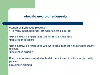
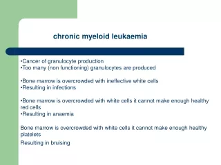
CHRONIC MYELOID LEUKAEMIAS
The World Health Organization (WHO) classification assigns some chronic myeloid leukaemias to a myeloproliferative category and others, in which there are also dysplastic features, to a myeloproliferative/myelodysplastic category