Download
1 / 38
380 likes | 396 Views
Explore the three main functions of the nervous system, sensory input, integration, and motor output, as well as the cells that make up the nervous system. Understand how reflexes work and the role of supporting cells.

E N D
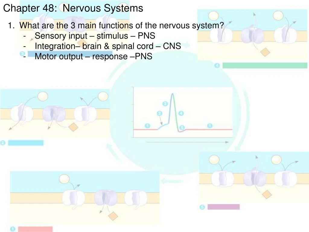
Chapter 48: Nervous Systems • What are the 3 main functions of the nervous system? • Sensory input – stimulus – PNS • Integration– brain & spinal cord – CNS • Motor output – response –PNS
Sensory input Integration Sensor Motor output Effector Peripheral nervoussystem (PNS) Central nervoussystem (CNS) Figure 48.3 Overview of information processing by nervous systems Protected by bone
Chapter 48: Nervous Systems • What are the 3 main fcns of the nervous system? • Sensory input – stimulus – PNS • Integration– brain & spinal cord – CNS • Motor output – response –PNS • 2. How does a reflex work?
Sensory neurons convey the information to the spinal cord. Sensors detect a sudden stretch in the quadriceps. The sensory neurons communicate with motor neurons that supply the quadriceps. The motor neurons convey signals to the quadriceps, causing it to contract and jerking the lower leg forward. 3 1 6 2 4 5 Cell body of sensory neuronin dorsal root ganglion Gray matter Sensory neurons from the quadriceps also communicate with interneuronsin the spinal cord. Quadricepsmuscle White matter Hamstringmuscle The interneurons inhibit motor neurons that supply the hamstring (flexor) muscle. This inhibition prevents the hamstring from contracting, which would resist the action of the quadriceps. Spinal cord(cross section) Sensory neuron Motor neuron The reflex is initiated by tapping the tendon connected to the quadriceps (extensor) muscle. Interneuron Figure 48.4 The knee-jerk reflex No brain involvement = faster response
Chapter 48: Nervous Systems • What are the 3 main fcns of the nervous system? • Sensory input – stimulus – PNS • Integration– brain & spinal cord – CNS • Motor output – response –PNS • How does a reflex work? • What cells make up the nervous system? • Neurons – functional unit of the nervous system • Supporting cells (glia) • Astrocytes, radial glia, oligodendrocytes, & Schwann cells • provide nutrition & support
Dendrites Cell body Nucleus Synapse Signal direction Axon Axon hillock Presynaptic cell Postsynaptic cell Myelin sheath Synapticterminals Figure 48.5 Structure of a vertebrate neuron Cell body – has nucleus Dendrites – bring signal to cell body Axon – takes signal away from cell body Axon hillock – cell body region where impulse is generated & axon begins Myelin – sheath that insulates axons made of supporting cells - PNS – Schwann cells secrete myelin - CNS – oligodendrocytes secrete myelin Synapse – junction between neurons or neuron & muscle or gland
Chapter 48: Nervous Systems • What are the 3 main fcns of the nervous system? • How does a reflex work? • What cells make up the nervous system? • Neurons – functional unit of the nervous system • Supporting cells (glia) • Astrocytes • regulate extracellular concentration of ions & neurotransmitters • Form tight junctions between cells that line capillaries of brain & • and spinal cord • Blood-brain barrier – restricts passage of substances into CNS • Can act as multipotent stem cells • Radial glia • Forms tracts for neurons to migrate in formation of neural tube • Oligodendrocytes & Schwann cells
Node of Ranvier Layers of myelin Axon Schwann cell Schwann cell Nodes of Ranvier Nucleus of Schwann cell Axon Myelin sheath 0.1 µm Figure 48.8 Schwann cells and the myelin sheath Node of Ranvier – space between Schwann cells on axon
Chapter 48: Nervous Systems Microelectrode –70 mV Voltage recorder Referenceelectrode • What are the 3 main fcns of the nervous system? • How does a reflex work? • What cells make up the nervous system? • What is the charge of a neuron? • -70 mV • WHY???
EXTRACELLULARFLUID CYTOSOL [Na+]150 mM + – [Na+]15 mM + – [K+]150 mM [K+]5 mM + – [Cl–]120 mM [Cl–]10 mM + – [A–]100 mM + – Plasmamembrane Figure 48.10 Ionic gradients across the plasma membrane of a mammalian neuron [A-] – DNA, RNA, proteins What happens when Na+ comes in & K+ leaves?
Inner chamber Outer chamber Inner chamber Outer chamber –92 mV +62 mV + – + – 150 mMNaCl 150 mMKCL 5 mMKCL 15 mMNaCl Cl– + – + – K+ Na+ Cl– Potassiumchannel + – Sodium channel + – Artificialmembrane (b) Membrane selectively permeable to Na+ (a) Membrane selectively permeable to K+ Figure 48.11 Modeling a mammalian neuron As K+ leaves, the cell loses (+) charge It becomes more (-) As Na+ enters, the cell gains (+) charge It becomes more (+)
Chapter 48: Nervous Systems • What are the 3 main fcns of the nervous system? • How does a reflex work? • What cells make up the nervous system? • What is the charge of a neuron? • How is neuron polarity altered?
Stronger depolarizing stimulus Stimuli Stimuli +50 +50 +50 Actionpotential 0 0 0 Membrane potential (mV) Membrane potential (mV) Membrane potential (mV) Threshold –50 –50 Threshold Threshold –50 Restingpotential Restingpotential Restingpotential Depolarizations Hyperpolarizations –100 –100 –100 0123456 0 1 2 3 4 5 0 1 2 3 4 5 Time (msec) Time (msec) Time (msec) (a) Graded hyperpolarizations produced by two stimuli that increase membrane permeability to K+. The larger stimulus producesa larger hyperpolarization. (b) Graded depolarizations produced by two stimuli that increase membrane permeability to Na+.The larger stimulus produces alarger depolarization. (c) Action potential triggered by a depolarization that reaches the threshold. Figure 48.12 Graded potentials and an action potential in a neuron Hyperpolarization K+ channels open Slight depolarization Na+ channels open More depolarization More Na+ enters Threshold achieved (-55 mV) LOTS of Na+ channels open NEURONS ARE ALL OR NONE!!
Chapter 48: Nervous Systems • What are the 3 main fcns of the nervous system? • How does a reflex work? • What cells make up the nervous system? • What is the charge of a neuron? • How is neuron polarity altered? • How is an action potential (nerve impulse) created?
+50 Actionpotential 0 Membrane potential (mV) Threshold Threshold –50 Resting potential –100 Time Extracellular fluid Activationgates Potassiumchannel Na+ 1 1 3 2 5 1 4 + + + + + + + + + + + + + + Plasma membrane – – – – – – – – – – – – – – Undershoot Cytosol Inactivationgate Sodiumchannel K+ Resting state Figure 48.13 The role of voltage-gated ion channels in the generation of an action potential
+50 Actionpotential Na+ Na+ 0 Membrane potential (mV) Threshold Threshold –50 K+ Resting potential –100 Time Depolarization Extracellular fluid Activationgates Potassiumchannel Na+ 2 1 3 2 5 1 4 1 + + + + + + + + + + + + + + + + + + + + + + Plasma membrane – – – – – – – – – – – – – – – – – – – – – – Cytosol Inactivationgate Sodiumchannel K+ Resting state Figure 48.13 The role of voltage-gated ion channels in the generation of an action potential
Na+ Na+ K+ Rising phase of the action potential +50 Actionpotential Na+ Na+ 0 Membrane potential (mV) Threshold Threshold –50 K+ Resting potential –100 Time Depolarization Extracellular fluid Activationgates Potassiumchannel Na+ 2 2 5 3 1 4 1 3 1 + + + + + + + + + + + + + + + + + + – – – – – – – – + + + + Plasma membrane – – – – – – – – + + + + + + + + – – – – – – – – – – – – – – Cytosol Inactivationgate Sodiumchannel K+ Resting state Figure 48.13 The role of voltage-gated ion channels in the generation of an action potential
Na+ Na+ Na+ Na+ K+ K+ Falling phase of the action potential Rising phase of the action potential +50 Actionpotential Na+ Na+ 0 Membrane potential (mV) Threshold Threshold –50 K+ Resting potential –100 Time Depolarization Extracellular fluid Activationgates Potassiumchannel Na+ 2 1 4 1 2 3 4 5 1 3 + + + + + + + + + + + + + + + + + + + + + + + + + + – – – – – – – – + + + + Plasma membrane – – – – – – – – + + + + + + + + – – – – – – – – – – – – – – – – – – – – – – Cytosol Inactivationgate Sodiumchannel K+ Resting state Figure 48.13 The role of voltage-gated ion channels in the generation of an action potential
Na+ Na+ Na+ Na+ K+ K+ Falling phase of the action potential Rising phase of the action potential +50 Actionpotential Na+ Na+ 0 Membrane potential (mV) Threshold Threshold –50 K+ Resting potential –100 Time Depolarization Na+ Na+ Extracellular fluid Activationgates Potassiumchannel Na+ 4 5 1 1 2 2 4 1 3 5 3 + + + + + + + + + + + + + + + + + + + + + + + + + + + + + + + + + + – – – – – – – – + + + + K+ Plasma membrane – – – – – – – – + + + + + + + + – – – – – – – – – – – – – – – – – – – – – – – – – – – – – – Undershoot Cytosol Inactivationgate Sodiumchannel K+ Resting state Figure 48.13 The role of voltage-gated ion channels in the generation of an action potential
Cytoplasmic Na+ binds to the sodium-potassium pump. EXTRACELLULAR FLUID [Na+] high [K+] low Na+ binding stimulates phosphorylation by ATP. Na+ Na+ Na+ Na+ Na+ ATP [Na+] low [K+] high P Na+ 1 5 2 4 3 6 ADP CYTOPLASM Na+ K+ is released and Na+ sites are receptive again; The cycle repeats. Phosphorylation causes the protein to change its conformation, expelling Na+ to the outside. Na+ Na+ K+ P K+ Extracellular K+ binds to the protein, triggering release of the Phosphate group. K+ Loss of the phosphate restores the protein’s original conformation. K+ K+ K+ P P i Figure 7.16 The sodium-potassium pump: a specific case of active transport Maintains charge of -70 mV. NOT THE SAME AS A Na+ or K+ channel.
Chapter 48: Nervous Systems • What are the 3 main fcns of the nervous system? • How does a reflex work? • What cells make up the nervous system? • What is the charge of a neuron? • How is neuron polarity altered? • How is an action potential (nerve impulse) created? • Why does an action potential only travel in 1 direction?
Axon Actionpotential – – + + + + + + An action potential is generated as Na+ flows inward across the membrane at one location. 1 + + – – – – – – Na+ – – – – – – + + – – + + + + + + Actionpotential The depolarization of the action potential spreads to the neighboring region of the membrane, re-initiating the action potential there. To the left of this region, the membrane is repolarizing as K+ flows outward. 2 K+ – – + + + + + + – – + – – – + – Na+ – – – – – – + + – – + + + + + + K+ Actionpotential The depolarization-repolarization process isrepeated in the next region of the membrane. In this way, local currents of ions across the plasma membrane cause the action potential to be propagated along the length of the axon. K+ – – – – + + + + 3 + + + + – – – – Na+ – – – + + – + + – – + + – – + + K+ Figure 48.14 Conduction of an action potential Domino analogy…
Schwann cell Depolarized region(node of Ranvier) Myelin sheath – –– – – – ++ + Cell body ++ ++ + Axon – – – ++ + – – – Figure 48.15 Saltatory conduction Depolarization jumps down the axon from node to node. Na+ & K+ channels are only found at the node of Ranvier. Action potentials can travel 120 m/sec
Chapter 48: Nervous Systems • What are the 3 main fcns of the nervous system? • How does a reflex work? • What cells make up the nervous system? • What is the charge of a neuron? • How is neuron polarity altered? • How is an action potential (nerve impulse) created? • Why does an action potential only travel in 1 direction? • How does a neuron communicate with another cell? • Chemical synapse • Signal changes from electrical chemical electrical
Postsynaptic cell Presynapticcell Na+ Synaptic vesiclescontainingneurotransmitter Neuro-transmitter K+ Presynapticmembrane Postsynaptic membrane Ligand-gatedion channel Voltage-gatedCa2+ channel Ca2+ Postsynaptic membrane 3 Synaptic cleft Ligand-gatedion channels 4 6 1 5 2 Figure 48.17 A chemical synapse
Chapter 48: Nervous Systems • What are the 3 main fcns of the nervous system? • How does a reflex work? • What cells make up the nervous system? • What is the charge of a neuron? • How is neuron polarity altered? • How is an action potential (nerve impulse) created? • Why does an action potential only travel in 1 direction? • How does a neuron communicate with another cell? • How does a single neuron interpret multiple inputs?
Terminal branch of presynaptic neuron Postsynaptic neuron E1 E1 E1 E1 E2 I Axonhillock Actionpotential Actionpotential Threshold of axon of postsynaptic neuron 0 Restingpotential Membrane potential (mV) –70 E1 E1 + E2 E1 E1 E1 E1 I E1 + I (c) Spatial summation (d) Spatial summationof EPSP and IPSP (a) Subthreshold, nosummation (b) Temporal summation Figure 48.18 Summation of postsynaptic potentials Axon hillock determines overall charge. If threshold is met then action potential is fired.
Na+ K+
Chapter 48: Nervous Systems • What are the 3 main fcns of the nervous system? • How does a reflex work? • What cells make up the nervous system? • What is the charge of a neuron? • How is neuron polarity altered? • How is an action potential (nerve impulse) created? • Why does an action potential only travel in 1 direction? • How does a neuron communicate with another cell? • How does a single neuron interpret multiple inputs? • Let’s look at some neurotransmitters….
Chapter 48: Nervous Systems • What are the 3 main fcns of the nervous system? • How does a reflex work? • What cells make up the nervous system? • What is the charge of a neuron? • How is neuron polarity altered? • How is an action potential (nerve impulse) created? • Why does an action potential only travel in 1 direction? • How does a neuron communicate with another cell? • How does a single neuron interpret multiple inputs? • Let’s look at some neurotransmitters…. • How is the nervous system organized?
Central nervous system (CNS) Peripheral nervous system (PNS) Brain Cranial nerves Spinal cord Ganglia outside CNS Spinal nerves Figure 48.19 The vertebrate nervous system
Gray matter White matter Ventricles Figure 48.20 Ventricles, gray matter, and white matter Gray matter – dendrites, unmyelinated axons & neuron cell bodies White matter – myelinated axons (myelin = white) Ventricles – filled with CSF (cerebrospinal fluid)
Peripheral nervous system Somatic nervous system Autonomic nervous system Sympathetic division Parasympathetic division Enteric division Figure 48.21 Functional hierarchy of the vertebrate peripheral nervous system
Parasympathetic division Sympathetic division Action on target organs: Action on target organs: Dilates pupil of eye Constricts pupil of eye Location of preganglionic neurons: brainstem and sacral segments of spinal cord Location of preganglionic neurons: thoracic and lumbar segments of spinal cord Inhibits salivary gland secretion Stimulates salivary gland secretion Sympathetic ganglia Neurotransmitter released by preganglionic neurons: acetylcholine Constricts bronchi in lungs Relaxes bronchi in lungs Neurotransmitter released by preganglionic neurons: acetylcholine Cervical Accelerates heart Slows heart Inhibits activity of stomach and intestines Thoracic Stimulates activity of stomach and intestines Location of postganglionic neurons: in ganglia close to or within target organs Location of postganglionic neurons: some in ganglia close to target organs; others in a chain of ganglia near spinal cord Inhibits activity of pancreas Stimulates activity of pancreas Stimulates glucose release from liver; inhibits gallbladder Stimulates gallbladder Lumbar Neurotransmitter released by postganglionic neurons: acetylcholine Neurotransmitter released by postganglionic neurons: norepinephrine Stimulates adrenal medulla Promotes emptying of bladder Inhibits emptying of bladder Promotes erection of genitalia Promotes ejaculation and vaginal contractions Sacral Synapse Figure 48.22 The parasympathetic and sympathetic divisions of the autonomic nervous system Rest & digest Fight or flight
Please take your test folder from the table by the door. • Crash Course Videos: • #26, 32, 33 • Bozeman Science Videos: • #18, 21, 22, 23, 36, 38, 41, 45
Unit 9 Test • 1) Put your L. Log in the blue bin. • 2) Put your cans (x3) in the green bin and sign the can log. • Animal Test cans due Friday, 4/17 • If you turn in 6 cans, you’ll receive can credit for BOTH the Animal Test & the Plant Test • 3) I’ll give you a used scantron. You’ll use the GREEN side today. • Mark your KEY ID (A or B) • 4) Write your name on EVERYTHING except the released AP ? Sheet.
Unit 9 Test Results • Average: 12 out of 18 • Range: 2 – 18 • Most-missed questions: • A #10, B #1 • A #11, B #11 • A #5, B #2 • Need 7.5/10 total score on L. Logs to be eligible for test corrections. • Test corrections are due on THURSDAY.


