Download
1 / 85
850 likes | 933 Views
PNS & Senses Ch. 14 & 15. Lindsey Bily Austin High School Anatomy & Physiology. Spinal Nerves. 31 pairs of nerves that are connected to the spinal cord. They are not named, just numbered based on where they emerge from the spinal cavity.

E N D
PNS & SensesCh. 14 & 15 Lindsey Bily Austin High School Anatomy & Physiology
Spinal Nerves • 31 pairs of nerves that are connected to the spinal cord. • They are not named, just numbered based on where they emerge from the spinal cavity. • The lower end of the spinal cord and its nerves looks like a horse’s tail. Called cauda equina (latin: horse’s tail).
Spinal Nerves • All spinal nerves contain both sensory and motor fibers, mixed nerves. • A plexus is where several fibers emerging from the spinal cord come together to form nerves. Plexus is latin for “Braid”. • Cervical plexus • Brachial plexus • Lumbosacral plexus
Plexus • Damage to one spinal nerve does not mean complete loss of function to a region of the body.
Cervical Plexus • Found deep within the neck. • Come from C1 through C5 vertebrae. • Nerves innervate the muscles and skin of the neck, upper shoulders, and part of the head. • Phrenic Nerve: innervates the diaphragm and exits the cervical plexus.
Brachial Plexus • Found deep within the shoulder. • Spinal nerves come from C5-T1 vertebrae and pass beneath the clavicle and toward the upper arm. • Innervate the lower part of the shoulder and entire arm.
Lumbosacral Plexus • Nerves join from the lumbar and sacral vertebrae. • Major nerves that emerge are the femoral nerve and the sciatic nerve.
Sciatic Nerve • Runs all the way down the back of the leg. • Supplies nearly all the skin of the posterior thigh, and leg and foot muscles. • Sciatica: pain in the nerve. It is very common and very painful.
Dermatomes and Myotomes • Areas on the skin or muscle that are innervated by sensory fibers of a given nerve. • Used to detect spinal cord or nerve abnormalities.
Herpes Zoster (Shingles) • Caused by the varicella zoster virus of chickenpox. • Usually the virus traveled through a cutaneous nerve and remained dormant for years. • May be reactivated during times of stress, low immunity or in the elderly. • Usually affects the skin of only one dermatome.
Cranial Nerves • 12 pairs of nerves that connect to the bottom of the brain, mostly on the brainstem. • Remember the mnemonic device. You will sound really smart in college if you already know this • On Old Olympus Towering Tops AFinn And German View Some Hops
Cranial Nerves Olfactory Optic Oculomotor Trochlear Trigeminal Abducens Facial Auditory (Vestibulocochlear) Glossopharyngeal Vagus Spinal Accessory Hypoglossal
Olfactory Nerve (I) • Olfactory: receptors are in the nose. Allow you to smell.
Optic Nerve (II) Optic: receptors are in the retina of the eye and include the optic nerves which come together at the optic chiasma to form the optic tract. The tract terminates in the thalamus and meets up with new fibers that go to the occipital lobe.
Oculomotor Nerve (III) • Fibers originate from cells in the midbrain. • Extend to the external eye muscles. • Autonomic fibers go to the intrinsic muscles of the eye and control the amount of light entering the eye and focusing.
Trochlear Nerve (IV) • Motor fibers originate from cell bodies in the midbrain and extend to the superior oblique muscles of the eye. Lets you move your eye.
Trigeminal Nerve (V) • Named so because they split off into 3 large branches. (tri-”three”; geminal-”pair”) • Opthalamic nerve • Maxillary nerve • Mandibular nerve
Abducens Nerve (VI) • Got its name because it abducts the eye. • Fibers originate in the pons and extend to the lateral rectus muscle of the eye.
Facial Nerve (VII) • Has motor fibers that arise from the pons and move the superficial muscles of the face and scalp. • Efferent autonomic fibers go to the tear ducts and the salivary glands. • Sensory fibers in the taste buds.
Facial Nerve (VII) • Bell’s Palsy: form of facial paralysis due to a problem with the facial nerve. • Can be caused by a stroke, brain tumor, or Lyme Disease. • Half your face appears to droop, your smile is one-sided, and your eye resists closing.
Auditory (Vestibulocochlear) Nerve (VIII) • Has two divisions (vestibular nerve and cochlear nerve). They are both sensory. • Vestibular nerve lets us know if we are balanced. • Cochlear nerve allows us to hear.
Glossopharyngeal Nerve (IX) • Sensory and motor fibers that innervate the tongue and pharynx (throat) and has a role in blood pressure.
Vagus Nerve (X) • Vagus means “wanderer”. Named this because it has so many branches. • Sensory fibers innervate the pharynx, larynx, trachea, heart, bronchi, esophagus, stomach, small intestine and gall bladder. • Motor fibers allow us to swallow. • Autonomic fibers control our heart rate and other vital activities.
Spinal Accessory Nerve (XI) • Named because it is an “accessory” to the Vagus nerve. • Has motor fibers that originate in the medulla oblongata and go to the thoracic and abdominal organs and the pharynx and larynx. • Can be a source of neck pain.
Hypoglossal Nerve (XII) • Means “under the tongue”. • Allows the tongue to move.
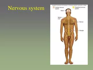
Autonomic Nervous System • Regulates all activities that we cannot control on our own. (involuntary). • Major function is to regulate heartbeat, smooth muscle contraction, and secretion by glands to maintain homeostasis. • 2 Efferent Divisions • Sympathetic division (fight or flight) • Parasympathetic division (rest and repair)
Autonomic Neurotransmitters and Receptors • Autonomic neurons release norepinephrine (NE) or Acetylcholine (ACH). • After they are released, they are either taken back up by the neuron that released them, or enzymes break them down.
Norepinephrine • Released by sympathetic division. • Causes… • Binds to beta receptors on cardiac muscle to speed up heart rate. • Constricts skin blood vessels • Dilates skeletal muscle blood vessels • Constricts abdominal blood vessels • Constricts blood vessels in the external genitalia • Dilates bronchioles in lungs • Decreases peristalsis • Relaxes the bladder • Dilates the pupil • Goosebumps • Increased sweat • Decreases production of saliva • Increases liver production of glucose
Beta Blockers • Drugs that bind to beta receptors. • They block the binding of NE and epinephrine (released by the adrenal glands). • They prevent increased heart rate that is caused by the sympathetic division.
Acetylcholine • Released by parasympathetic division. • Causes… • heart rate to decrease • heart blood vessels to dilate. • blood vessels in the external genitalia to dilate • Increased peristalsis (contraction of digestive tract). • Contraction of bladder • Constriction of pupil • Increased secretion of tears • Increased secretion of saliva • Increased secretion of pancreatic juice and insulin
Sense Organs • We have millions of sense organs! • Receptors: general sense organs. Produce general senses (touch, temperature, pain). • Vision, hearing, balance, taste and smell are special sense organs.
Receptors • Respond to stimuli by converting them to nerve impulses. • Different types of receptors respond to different stimuli. (ex. heat receptors cannot sense light or pain) • When a stimulus acts on a receptor, a receptor potential is created. If the stimulus is strong enough, it can create and action potential. • We then interpret the sensation in the brain and/or spinal cord. • Adaptation: the magnitude of the receptor potential decreases over time in response to a constant stimulus. (You don’t feel your clothes a few minutes after you put them on).
Distribution of Receptors • Millions of receptors all over the parts of our body. But not uniform. • For instance, the fingertips have many more touch receptors than does the skin on the back.
Classification of Receptors • Can be classified by location, stimulus detected, or structure. • Location • Exteroreceptors: near the body surface (pressure, touch, pain, temperature) • Visceroreceptors: located internally, often within an organ (pressure, stretch, chemical changes) • Proprioreceptors: more specialized and in skeletal muscle, joint capsules and tendons. Tells us information about body movement, orientation in space and muscle stretch.
Classification of Receptors • Stimulus Detected • Mechanoreceptors: mechanical stimuli “deform” or change the position of the receptor (pressure receptor). • Chemoreceptors: activated by the change in amount or concentration of chemicals (pH, glucose levels) • Thermoreceptors: changes in temperature • Nociceptors: intense stimuli of any type that results in tissue damage that causes pain. • Photoreceptors: found only in the eye and responds to light stimuli.
Classification of Receptors • Structure • Free nerve endings: simplest and most common. • Nociceptors (pain) • Merkel disks (light pressure) • Root hair plexuses (hair movement) • Encapsulated nerve endings: have a connective tissue capsule that surrounds their terminal or dendritic end. • Meissner’s corpuscle (light pressure in the epidermis of hairless skin ex. Nipples, fingertips, lips) • Krause’s end bulb (touch and low vibration in mucous membranes of eye, lips, tongue) • Pacinian corpuscle: (deep pressure, stretch, high vibration in dermis and joints) • Ruffini’s corpuscle: (crude and persistent touch in dermis of skin Ex. Steering the wheel of a car) • Muscle spindles: (stretch in skeletal muscle) • Golgi tendon receptors: (muscle tension in the junction between muscle and tendon).
Sense of Smell • Olfactory receptors are located in the most superior portion of the nasal cavity. This is why we have to sniff to smell really delicate odors. • We have olfactory epithelium that has epithelial yellow support cells, basal cells, and bipolar receptor neurons. • The receptors are chemoreceptors. Gas molecules or chemicals dissolved in the mucus bind to the receptors to cause receptor potentials.
Sense of Smell • Most humans can distinguish between hundreds of odors. Some several thousand odors. • Well known primary scents: • Putrid, floral, peppermint, and musky odors • Combinations of two or more produce the wide array of odors that we know. • It appears that we become adapted to odors quickly due to inhibition of action potentials by granule cells in the nose as opposed to the neurons adapting.
Olfactory Pathway Path of action potential… Receptor nerve olfactory tract thalamus olfactory centers of the brain Smell can produce long lasting memories from childhood to adult. Smells are ultimately processed in the temporal lobes. However, axons go to limbic system and hippocampus.
Sense of Taste • Most of the taste buds are on the tongue, but we do have some in the lining of the mouth and soft palate. • Papillae are small elevated projections on the tongue. • Fungiform, circumvallate, and foliate papillae all contain taste buds. • Threadlike filiformpapillae don’t have taste buds but let us detect food texture and “feel”.
Taste • There is no “tongue map” like originally thought. All tastes can be detected in all areas of the tongue that contain taste buds. • Taste buds contain chemoreceptors that detect chemicals dissolved in saliva. • Primary taste sensations: • Sour, sweet, bitter, salty (umami, metallic) Other flavors we taste are combinations of these.
Taste • Taste receptors are very sensitive and tend to fatigue easily like olfactory receptors. • Pathway… Receptor in taste bud facial nerve (if in front 2/3 of tongue/glossopharyngeal nerve (if in back 1/3)/vagus nerve (from taste buds located in the pharynx and epiglottis) medulla oblongata thalamus parietal lobe of cerebrum
Hearing and Balance • The ear allows us to hear, but also to balance. • The “trigger” for this is caused by activation of mechanoreceptors called hair cells. • Sound waves and movement cause the forces to change the shape of the receptors.
External Ear • Has two parts: pinna (on the side of the head) and the external auditory meatus (ear canal) • The ear canal is about 3 cm long and ceruminous glands secrete cerumen (wax). • Tympanic membrane (eardrum) separates the ear canal from the middle ear.