Download
1 / 29
290 likes | 406 Views
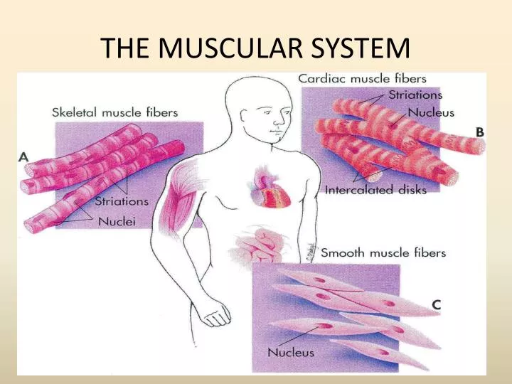
THE MUSCULAR SYSTEM. What types of muscles can be found in the body?. Smooth Muscle. Cardiac Muscle . Skeletal Muscle Fibers. Figure 6.1. Pectoralis major Draws arm forward and toward the body. Trapezius Lifts shoulder blade Braces shoulder Draws head back. Deltoid Raises arm.

E N D
Figure 6.1 • Pectoralis major • Draws arm forward • and toward the body • Trapezius • Lifts shoulder blade • Braces shoulder • Draws head back • Deltoid • Raises arm • Biceps brachii • Bends forearm at elbow • Triceps brachi • Straightens forearm • at elbow • Rectus abdominus • Compresses abdomen • Bends backbone • Compresses chest cavity • Latissimus dorsi • Rotates and draws • arm backward and • toward body • External oblique • Lateral rotation of trunk • Compresses abdomen • Gluteus maximus • Extends thigh • Rotates thigh laterally • Gastrocnemius • Bends lower leg at • knee • Bends foot away from • knee
Skeletal muscle microstructure. Myofibril Z-line Z-line Sarcomere A closer view of a section of a myofibril showing that it is composed of sarcomeres joined end to end at the Z-line. Myosin Actin An electron micrograph cross section of a sarcomere in a region that contains both actin and myosin. Thin filament (actin) Thick filament (myosin) Sarcomeres contain thin filaments of actin that attach to the Z-lines and thicker filaments of myosin that span the gap between actin molecules. A transmission electron micrograph ( 11,300) of a longitudinal section of a sarcomere. The rounded red objects are mitochondria.
Activity 1: Skeletal Muscle • Has striated cells with multiple nuclei • Occurs in the muscles attached to skeleton • Functions in voluntary movement of body • Examine figure 1. • Identify the long, multinucleated fibers arranged in a parallel fashion. How do you know the muscle fibers are striated?
Activity 1 • Draw a sarcomere. • Identify and label the following structures: • Z-lines • Thick and Thin filaments
Activity 2. • Examine the model of the neuromuscular junction. • Examine the branching of the motor neuron to several muscle cells. • Identify the motor neuron, synaptic cleft, sarcoplasmic reticulum and T- tubules. • Draw the neuromuscular junction.
Figure 6.6 1 The release of acetylcholine at the neuromuscular junction causes an electrical impulse to be generated in the muscle cell plasma membrane Motor neuron Acetylcholine 2 The electrical impulse ( ) is carried to the cell’s interior by the T tubules Electrical impulse Ca2 T tubule Sarcoplasmic reticulum 3 The electrical impulse triggers the release of Ca2 from the sarcoplasmic reticulum Muscle cell plasma membrane Myofibrils Z-line
Questions • How do a motor neuron and a skeletal muscle fiber join at a neuromuscular junction? • What events occur at this neuromuscular junction?
Activity 3 • Examine the figure of a muscle cross section. • Identify the following structures: tendon, fascicles, perimysium, epimysium. • Draw and label whole muscle.
Activity 4: observing the gross anatomy of skeletal muscles.
Figure 6.1 • Pectoralis major • Draws arm forward • and toward the body • Trapezius • Lifts shoulder blade • Braces shoulder • Draws head back • Serratus anterior • Helps raise arm • Contributes to pushes • Draws shoulder blade forward • Deltoid • Raises arm • Biceps brachii • Bends forearm at elbow • Triceps brachi • Straightens forearm • at elbow • Rectus abdominus • Compresses abdomen • Bends backbone • Compresses chest cavity • Latissimus dorsi • Rotates and draws • arm backward and • toward body • External oblique • Lateral rotation of trunk • Compresses abdomen • Gluteus maximus • Extends thigh • Rotates thigh laterally • Adductor longus • Flexes thigh • Rotates thigh laterally • Draws thigh toward body • Hamstring group • Draws thigh backward • Bends knee • Sartorius • Bends thigh at hip • Bends lower leg at knee • Rotates thigh outward • Gastrocnemius • Bends lower leg at • knee • Bends foot away from • knee • Quadriceps group • Flexes thigh at hips • Extends leg at knee • Achilles tendon • Connects • gastrocnemius • muscle to heel • Tibialis anterior • Flexes foot toward knee
Activity 4 • Complete table 9.4
Activity 5: Contraction of Skeletal Muscles • Work in pairs • Obtain a gripper • Read the directions • Complete questions 1-3.
Due Date • March 11-14
Introduction Function of the muscular system Types of muscles and functions Basic structure of skeletal muscles Skeletal muscles work in pairs Name and actions of skeletal muscles Leave procedures out
Results Include your results from each activity. Drawing, label your drawings see the following example. Complete table or box. Label the table and include a description of the table or box.
Figure 1. Motor neuron anatomy. Neurons are cells specialized to conduct nerve impulses.
Conclusion • Include motor neuron and motor unit. • Include all the components of a neuromuscular junction. How do a motor neuron and skeletal muscle fiber join at a neuromuscular junction? What events occur at the neuromuscular junction? What is the function of ACh? • Include the microscopic anatomy of a muscle fiber • Describe the sarcomere(include all the components in a sarcomere) • Describe the contraction cycle. • What are the roles played by calcium and ATP during muscular contraction? • Explain cross-bridge, power stroke and the sliding filament model. • What is the function of ATPase? • Isometric and Isotonic contractions • Complete the questions page 113 and 114.