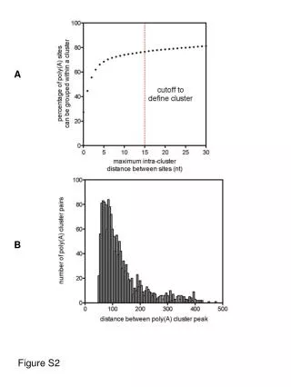
Download
1 / 1
10 likes | 72 Views
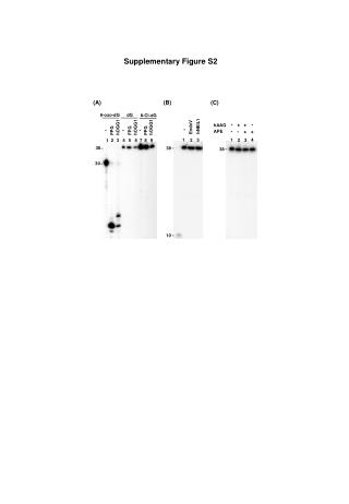
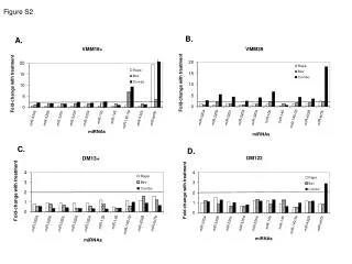
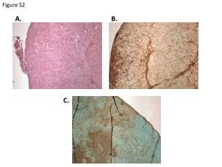
C. B. A. 1 2 3. 1 2 3. M 1 2 3. KD. 175. 175. 46. WB blot. CB stained gel. WB blot. Figure S2.

E N D
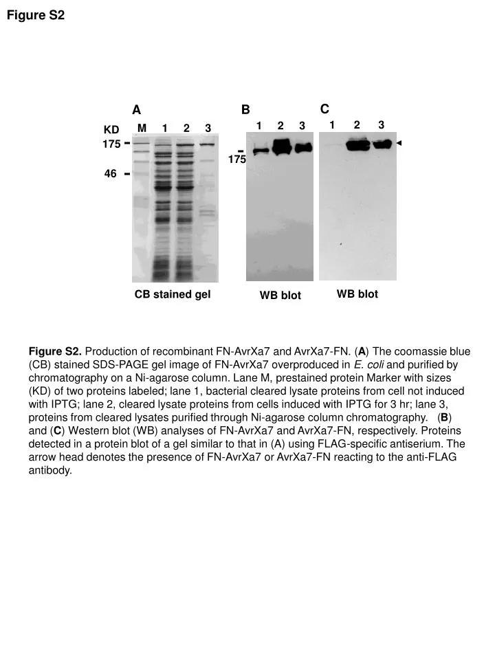
C B A 1 2 3 1 2 3 M 1 2 3 KD 175 175 46 WB blot CB stained gel WB blot Figure S2 Figure S2. Production of recombinant FN-AvrXa7 and AvrXa7-FN. (A) The coomassie blue (CB) stained SDS-PAGE gel image of FN-AvrXa7 overproduced in E. coli and purified by chromatography on a Ni-agarose column. Lane M, prestained protein Marker with sizes (KD) of two proteins labeled; lane 1, bacterial cleared lysate proteins from cell not induced with IPTG; lane 2, cleared lysate proteins from cells induced with IPTG for 3 hr; lane 3, proteins from cleared lysates purified through Ni-agarose column chromatography. (B) and (C) Western blot (WB) analyses of FN-AvrXa7 and AvrXa7-FN, respectively. Proteins detected in a protein blot of a gel similar to that in (A) using FLAG-specific antiserium. The arrow head denotes the presence of FN-AvrXa7 or AvrXa7-FN reacting to the anti-FLAG antibody.