Download
1 / 37
390 likes | 575 Views
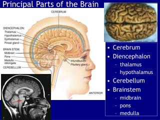
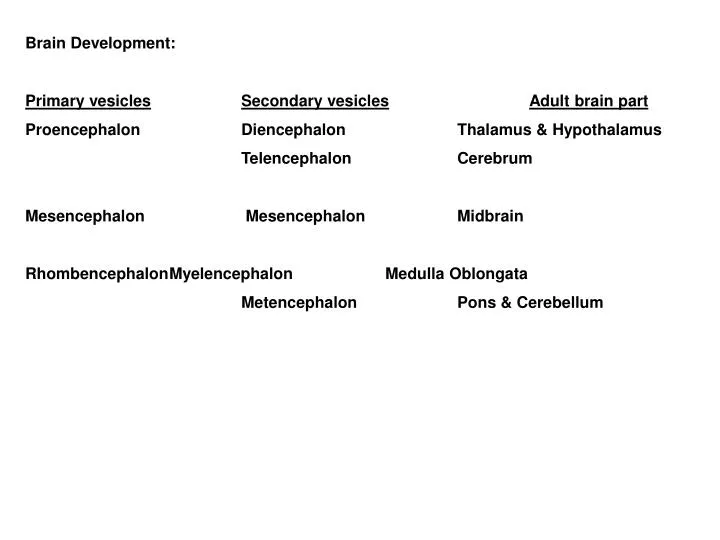
Brain Development: Primary vesicles Secondary vesicles Adult brain part Proencephalon Diencephalon Thalamus & Hypothalamus Telencephalon Cerebrum Mesencephalon Mesencephalon Midbrain Rhombencephalon Myelencephalon Medulla Oblongata Metencephalon Pons & Cerebellum.

E N D
Brain Development: Primary vesiclesSecondary vesiclesAdult brain part Proencephalon Diencephalon Thalamus & Hypothalamus Telencephalon Cerebrum Mesencephalon Mesencephalon Midbrain Rhombencephalon Myelencephalon Medulla Oblongata Metencephalon Pons & Cerebellum
Cerebrospinal fluid (CSF) - formed in choroid plexes of 4 ventricles: • 2 lateral - one in each hemisphere • 3rd - interventricular foramen connect with 3rd w/ both lateral vent. • 4th - cerebral aqueduct connects 3rd and 4th vent. • Three openings (apertures) from 4th into subarachnoid space: • 2 lateral, 1 median • Returns to blood via arachnoid villa • Internal hydrocephallus • External hydrocephallus • Blood-CSF barrier • Blood brain barrier (BBB)
Medulla: Ascending and descending tracts Inferior cerebellar peduncles - medulla to cerebellum Pyramids Decussation of pyramids Reflex centers (nuclei) Cardiac - CAC CIC Medullary rhythmicity - respirations Vasomotor - blood vessel diameter Cranial nerves VIII* - XII
Pons: • Ascending and descending tracts • Middle cerebellar peduncles - pons to cerebellum • Nuclei: • Pneumotaxic - respiration • Apneustic " • Cranial nerves V – VIII*
Midbrain: Cerebral peduncles - connect upper brain w/ brain stem and SC Superior cerebellar peduncles - midbrain to cerebelum Cerebral aqueduct Corpora Quadrigemina: Superior colliculi – reflex center for eyes, head, neck in response to visual stimuli Inferior colliculi – reflex center for head, trunk in response to auditory stimuli Nuclei: Substantia Nigra – control subconscious myo activity Red Nuclei – coordinates myo activity w/ basal ganglia, cerebellum Cranial nerves III – IV
Reticular Formation: gray matter among white matter in medulla, pons, midbrain sensory and motor functions RAS – consciousness and arousal Hypothalamus: Involved with homeostasis, hunger, thirst, etc. Secondary controller of emotional behavior Thalamus: Receives and interprets all sensory input (Cranial Nerve I -Olfactory). Relays information to sensory cortex of brain.
Cerebrum: Outer layer - cerebral cortex (gray), gyri Inner layer - white Longitudinal fissure - hemispheres (lobes), falx cerebri, corpus callosum Central sulcus - separates frontal & parietal Lateral cerebral sulcus - separates frontal & temporal Parieto -occipital sulcus - separates parietal & occipital Transverse fissure - separates cerebrum & cerebellum Three sets of fibers: Association - w/i same hemi Commissural - between corresponding gyri in each hemi Projection - Ascending and descending tracts Basal ganglia - paired masses of gray matter Corpus Striatum Caudate Nucleus - controls large subconscious movement Lentiform Nucleus Putamen - same as Caudate Globus Pallidus - regulates myo tone for specific movements Limbic system - gray matter, primary controller of emotions
Cerebral cortex: Motor areas Primary - controls groups of skeletal myo Broca's - control myo for speech Sensory areas Primary somesthetic - locate point of stimuli Association areas Premotor - generates impulses to control complex, sequential learned movements (writing) Somesthetic association - integrates/interprets Wernicke's - comprehension written & spoken language. Fibers connect w/ Broca's Cerebral asymetry L hemi - verbal, math, analytical, etc. R hemi - non-verbal, music, spatial, etc.
Cerebellum: Transverse fissure (Tentorium Cerebelli – cranial meninges in TF) Vermis - constricted portion, divides into hemi (falx cerebelli) Hemi divided into lobes Anterior - controls large subconscious skeletal movement Posterior - same Flocculonodular - equilibrium, posture Three sets of fibers Inferior cerebellar peduncles Middle " " Superior " "
Levels of Motor Control Low levels controlled by reflex arcs. Complex levels walking/swimming (FAPs) Segmental - CPGs Projection - command nuerons Program/instruction - Cerebellum and basal ganglia