Download
1 / 2
20 likes | 25 Views
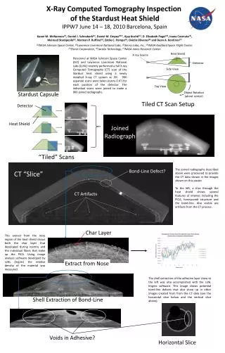
En'Urga Inc. has a full X-Ray tomography system which can be used to perform a non-destructive evaluation of plastic components. In addition, the system can provide soot mass concentrations in diesel particulate filters<br><br>

E N D
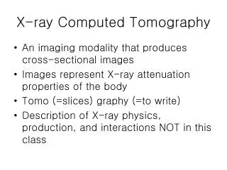
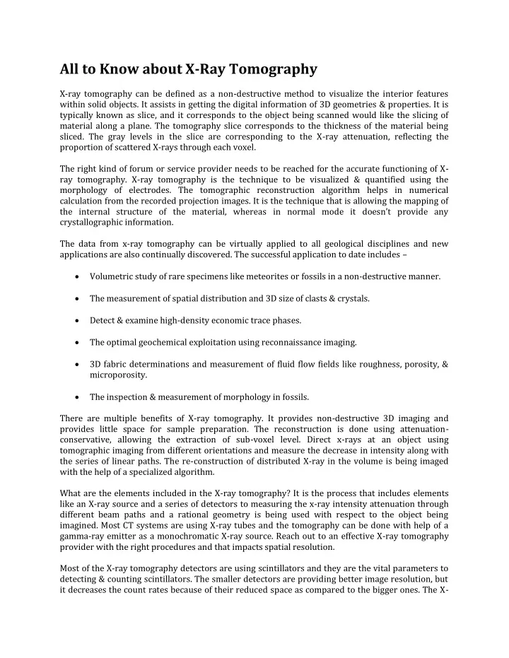
All to Know about X-Ray Tomography X-ray tomography can be defined as a non-destructive method to visualize the interior features within solid objects. It assists in getting the digital information of 3D geometries & properties. It is typically known as slice, and it corresponds to the object being scanned would like the slicing of material along a plane. The tomography slice corresponds to the thickness of the material being sliced. The gray levels in the slice are corresponding to the X-ray attenuation, reflecting the proportion of scattered X-rays through each voxel. The right kind of forum or service provider needs to be reached for the accurate functioning of X- ray tomography. X-ray tomography is the technique to be visualized & quantified using the morphology of electrodes. The tomographic reconstruction algorithm helps in numerical calculation from the recorded projection images. It is the technique that is allowing the mapping of the internal structure of the material, whereas in normal mode it doesn’t provide any crystallographic information. The data from x-ray tomography can be virtually applied to all geological disciplines and new applications are also continually discovered. The successful application to date includes – Volumetric study of rare specimens like meteorites or fossils in a non-destructive manner. The measurement of spatial distribution and 3D size of clasts & crystals. Detect & examine high-density economic trace phases. The optimal geochemical exploitation using reconnaissance imaging. 3D fabric determinations and measurement of fluid flow fields like roughness, porosity, & microporosity. The inspection & measurement of morphology in fossils. There are multiple benefits of X-ray tomography. It provides non-destructive 3D imaging and provides little space for sample preparation. The reconstruction is done using attenuation- conservative, allowing the extraction of sub-voxel level. Direct x-rays at an object using tomographic imaging from different orientations and measure the decrease in intensity along with the series of linear paths. The re-construction of distributed X-ray in the volume is being imaged with the help of a specialized algorithm. What are the elements included in the X-ray tomography? It is the process that includes elements like an X-ray source and a series of detectors to measuring the x-ray intensity attenuation through different beam paths and a rational geometry is being used with respect to the object being imagined. Most CT systems are using X-ray tubes and the tomography can be done with help of a gamma-ray emitter as a monochromatic X-ray source. Reach out to an effective X-ray tomography provider with the right procedures and that impacts spatial resolution. Most of the X-ray tomography detectors are using scintillators and they are the vital parameters to detecting & counting scintillators. The smaller detectors are providing better image resolution, but it decreases the count rates because of their reduced space as compared to the bigger ones. The X-
ray orientation methods are based on transmitted X-rays and high-energy radiation is needed to achieve the desired parameters. Multiple detectors are used by high-energy diffraction microscopy to determine the beam’s origins. The process makes the three-dimensional orientation of maps possible and subjects the sample to external stimulus. Source: https://www.enurga.com/xraytomography.htm