Download

1 / 102
1.07k likes | 1.32k Views
Urinary Bladder: Congenital Anomalies trauma, cystitis, tumors, urethral trauma. Dr.Amit Gupta Associate Professor Dept. of Surgery. Anatomy. The urinary bladder is a hollow, muscular , and distensible (or elastic) organ that sits on the pelvic floor .

E N D
Urinary Bladder: Congenital Anomalies trauma, cystitis, tumors, urethral trauma Dr.Amit Gupta Associate Professor Dept. of Surgery
Anatomy • The urinary bladder is a hollow, muscular, and distensible (or elastic) organ that sits on the pelvic floor . • It is the organ that collects urine excreted by the kidneys prior to disposal by urination. Urine enters the bladder via the ureters and exits via the urethra. • In males, the base of the bladder lies between the rectum and the pubic symphysis. It is superior to the prostate, and separated from the rectum by the rectovesical pouch. • In females, the bladder sits inferior to the uterus and anterior to the vagina. It is separated from the uterus by the vesicouterine pouch.
Embryology • The urogenital sinus is formed by the division of the cloaca by the uro-rectal septum • The uro-genital sinus may be divided into three component parts. • cranial portion which is continuous with the allantois and forms the bladder proper. • The pelvic part of the sinus forms the prostatic urethra and epithelium as well as the membranous urethra and bulbo urethral glands in the male and the membranous urethra and part of the vagina in females. • the caudal portion forms the penile urethra in males and the vestibule in females.
Embryology…….. • The bladder forms from the cranial end of the urogenital sinus. However, the trigone portion is formed by the caudal ends of the mesonephric ducts. As the bladder expands, the mesonephric ducts begin to become incorporated into the wall of the bladder dragging the ureters along with them. Further growth causes the ureters to eventually have their own opening into the bladder. • The bladder is initially continuous with the allantois. Over time, the allantois degenerates to form a cord-like structure, the urachus. The urachus goes from the umbilicus to the apex of the bladder and forms the median umbilical ligament which can be seen in adults. The medial umbilical ligaments may also be seen in adults on both sides of the median umbilical ligament, these are the vestigial remnants of the umbilical arteries.
Congenital Anomalies A major consideration with congenital abnormalities is that they tend to be multiple. All of these anomalies are infrequent or rare, and each condition occurs in both males and females Diagnosed in infancy or childhood Most are discovered in the evaluation of a urinary tract infection or, in the case of urachal anomalies, periumbilical drainage or redness.
Classification Urinary bladder anomalies are- • bladder diverticula • bladder ears • congenital hypoplasia of the bladder • megacystis • bladder agenesis • duplication anomalies of the bladder • bladder septa Urachal anomalies are urachal sinus urachal cyst urachal diverticulum patent urachus
Pathophysiology The embryologic cause is unknown. Bladder development occurs during the fifth to seventh week of gestational development. • Development depends upon many factors • mesenchymal differentiation • mesenchymal growth • urine production Bladder cycling, the process of sequential expansion and contraction, is important in the anatomic and physiologic development of the normal bladder.
Bladder Diverticulum Bladder diverticula are herniations of the bladder mucosa through bladder wall musculature (detrusor muscle). • Diverticular size can vary • Diverticula can be wide or narrow mouthed.(The size of diverticular openings has functional implications because narrow-mouthed diverticula often empty poorly). • Stasis of urine within diverticula can also lead to stone formation or epithelial dysplasia. • may cause ureteral obstruction,bladder outlet obstruction or vesicoureteral reflux
Bladder Diverticulum…………… MC Site lateral and superior to the ureteral orifices. [dome of the bladder, in disorders as bladder outlet obstruction (ie, posterior urethral valves) or Eagle Barrett syndrome (prune belly syndrome)]. Congenital or acquired. Congenital deficiency or weakness in the Waldeyer fascial sheath has been implicated as a cause. Solitary located at the junction of the bladder trigone and detrusor Congenital diverticula are usually removed surgically
Bladder ears lateral protrusions of the bladder through the internal inguinal ring and into the inguinal canal. Bladder ears are often observed during voiding cystourethrography (VCUG) or intravenous pyelography (IVP), when the bladder is filled to capacity. Bladder ears have also been seen on CT body imaging. No treatment is necessary Knowledge of this entity is important to surgeons during inguinal herniorrhaphy because occasional reports have been made of partial or near total cystectomy performed under the mistaken notion that this was a large hernia sac.
Bladder agenesis • Rare • generally incompatible with life. • Ureters may enter into the urethra, vagina, Gartner duct cyst (female), prostatic urethra, rectum, or the patent urachus. • Associated hydroureteronephrosis and renal dysplasia (variable) are present. • Other associated anomalies include neurologic, orthopedic, hindgut, and other urogenital anomalies, such as renal agenesis and absence of the prostate, vagina, seminal vesicles, epididymis, or penis This condition requires urinary diversion and subsequent reconstruction
Megacystitis Megacystitis is an enlarged bladder, believed to be secondary to overfilling of the fetal bladder during development Common presentations- hydronephrosis. Febrile urinary tract infection high-grade vesicoureteral reflux Megacystitis can be observed in other conditions, such as posterior urethral valves, Ehlers-Danlos syndrome, urethral diverticulum, microcolon hypoperistalsis syndrome, sacral meningomyelocele, sacrococcygeal teratoma, and pelvic neuroblastoma. Clean intermittent catheterization
Bladder duplication • Rare • Complete (2 urethras exist) • Partial (the bladder joins distally into one common urethra) • The 2 halves of the bladder are on either side of the midline, with the corresponding ipsilateral ureter draining each bladder half. • Associated anomalies include duplication of the penis, vagina, uterus, lumbar vertebrae, and hindgut. fistulas may be present between the rectum, vagina, and urethra.
Urachal cyst • is a fluid-filled structure occurring in between the two obliterated ends of the urachus. • Most occur in the distal third of the urachus. • More commonly, the urachal cyst is detected in early childhood or adolescence. • Symptoms -infection ,inflammation,suprapubic mass, fever, pain, and bladder or irritative voiding symptoms. • (Staphylococcus aureus is the most common bacterial organism). • Death from intra-abdominal rupture has been reported.
Patent urachus Communication from the umbilicus to the bladder. continuous or intermittent drainage from the umbilicus. The tract may become inflamed, resulting in tenderness, periumbilical swelling, and serosanguinous or purulent discharge. May be associated with bladder outlet obstruction, such as posterior urethral valves. Correction of the obstruction may result in the spontaneous resolution of the patent urachus.
Urachal sinus derived from a persistently patent urachus Children may present with periumbilical tenderness wet umbilicus granulation tissue at the level of the umbilicus In many instances, these children have undergone multiple sliver nitrate cauterizations under the mistaken notion that this is simply granulation tissue after severance of the remnant umbilical cord. These can be observed in the first 4-8 weeks of life.
Ectopia Vesicae open to the outside and turned inside-out, so that its inside is visible at birth, protruding from the lower abdomen Symptoms- • Exposed posterior bladder wall • Short lower abdominal wall • Inguinal hernia • Separated pubic rami • Epispadias • Incomplete fusion of genital tubercles
Opening in abdominal wall • Urine excretion through opening in abdominal wall • Incontinence • Dilated ureters • Open pubic arch • Wide-set ischial bones • Constriction between ureter and bladder • Ureteral reflux
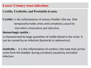
Cystitis • In womenis common due to a number of reasons : - short urethra - pregnancy - decreased estrogen production during menopause. • In men: mainly due to persistent bacterial infection of the prostate. • In both sexes: common risk factors are : - presence of bladder stone - urethral stricture - catheterization of the urinary tract - diabetes mellitus
Pathogenesis of cystitis Due to frequent irritation of the mucosal surfaces of the urethra and the bladder. Infection results when bacteria ascends to the urinary bladder . These bacteria are residents or transient members of the pereneal flora, and are derived from the large intestine flora. Toxins produced by uropathogens. Condition that create access to bladder are: - Sexual intercourse due to short urethral distance.
Pathogenesis of cystitis • Uncomplicated UTI usually occurred in non pregnant , young sexually active female without any structural or neurological abnormality • Risk factors : - Catheterization of the urinary bladder , instrumentation - structural abnormalities - obstruction • Hematogenous through blood stream ( less common) from other sites of infection
Etiologic agents • E.coli is the most common (90%) cause of cystitis. • Other Enterobacteria include ( Klebsiellapnumoniae, Proteus spp.) Other gram negative rods eg. P.aeroginosa. • Gram positive bacteria :Enterococcusfecalis, group B Strept. and Staphylococcus saprophyticus{ honeymoon cystitis}. • Candida species • Venereal diseases ( gonorrhea, Chlamydia) may present with cystitis. • Schistosomahematobiumin endemic areas.
Pathogens involved Uncomplicated UTI E. coli64% Enterobacteriaceae16% Enterococcusspp 20% Pseudomona spp <1% S. aureus <1% Special cases (S. epidermidis) S. saprophyticus Yeasts (catheter related result) Viruses (Adeno, Varicella) Chlamydia trachomatis Complicated UTI E.coli Enterobacteriaceae Pseudomonasspp Acinetobacterspp (judge often multiresistant strains) % is not possible to
Clinical presentation Symptoms usually of acute onset • Dysuria ( painful urination) • Frequency ( frequent voiding) • Urgency ( an imperative call for toilet) • Haematuria ( blood in urine) in 50% of cases. • Usually no fever.
Vaginitis (5%) Candida spp. T. vaginalis Cystitis (80%) E. coli, S. saprophyticus Proteus spp. Klebsiella spp. Urethritis (10-15%) C. trachomatis, N. gonorrhoeae H. simplex Other bacteria? Dysuria and frequency Non-infectious (<1%) Hypoestrogenism Functional obstruction Mechanical obstruction Chemicals
How to differentiate between cystitis and urethritis ? • Cystitis is of more acute onset • More sever symptoms • Pain, tenderness on the supra-pubic area. • Presence of bacteria in urine ( bacteriuria) • Urine cloudy, malodorous and may be bloody
Differential diagnosis ( types of cystitis) • Non-infectious cystitis such as: • Traumatic cystitis in women • Interstitial cystitis ( unknown cause, may be due to autoimmune attack of the bladder) • Eosinophilic cystitis due to S.hematobium • Hemorrahagic cystitis due to radiotherapy or chemotherapy.
Laboratory diagnosis of cystitis Specimen collection Microscopic examination Chemical screening tests
Recurrent cystitis 3 or more episodes of cystitis /year Requires further investigations such as Intravenous Urogram ( IVU) or ultrasound to detect obstruction or congenital deformity Cystoscopy required in some cases
Treatment of cystitis • Empiric treatment commonly used depending on the knowledge of common organism and sensitivity pattern • Treatment best guided by susceptibility pattern of the causative bacteria • Common agents: Ampicillin, Cephradine, Ciprofloxacin, Norfloxacin, Gentamicin or TRM-SMX.
Duration of treatment: 3 days for uncomplicated cystitis • 10-14 days for complicated and recurrent cystitis • Prophylaxis required for recurrent cases by Nitrofurantoin or TRM-SMX • Prevention : drinking plenty of water and prophylactic antibiotic
Aetiology • Smoking – 4x increased risk • Causes 50% of cases • Occupational – rubber/dye industry • Napthylamine, benzidine • Schistosomiasis, chronic infection • Medications – cyclofosfamid, fenacetin
Histology • 90-95% transitional-cellcarcinoma • 3% squamos-cell carcinoma • 2% adenocarcinoma • <1% small-cellcarcinoma • 99%primarytumors
Entities • 75-85% superficialbladdercancer pTa, pTis, pT1 • 10-15% muscle-invasive bladdercancer pT2, pT3, pT4 • 5% metastaticbladdercancer N+, M+
Presentation • Classically painless frank haematuria, sometimes intermittent • Frequent urination, urgency • symptoms with involvement of neighbouring organs,lymphoedema, pelvic pain
Examination • History • Physical examination • Urine examination / urinalysis, cultivation, cytology – can be only 60% sensitive/ • Ultrasound
Examination • Cystoscopy is mandatory • Biopsy or TURBT • Bimanual pelvic examination /before and after TURBT • Chest X-ray • IVU – no routinely, (5% chance upper tract involvement)