Download
1 / 26
310 likes | 503 Views
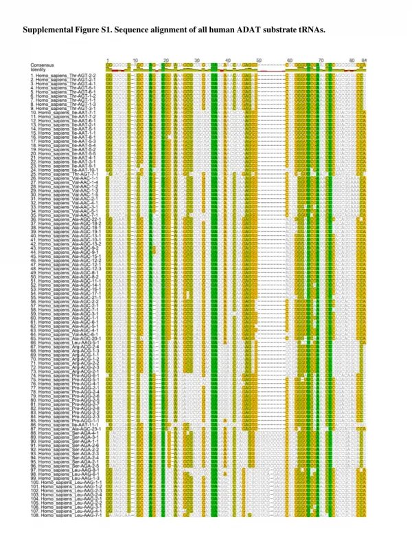
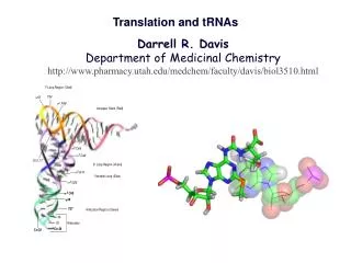
G. Structures of tRNAs. (a) tRNAs are 73~93 nucleotides long. (b) Contain several modified nucleotides. (c) The anticodon loop and the 3’ CCA of the acceptor stem. Two-step decoding process for translating nucleic acid sequences in mRNA into amino acid sequences in proteins.

E N D
Structures of tRNAs (a) tRNAs are 73~93 nucleotides long. (b) Contain several modified nucleotides. (c) The anticodon loop and the 3’ CCA of the acceptor stem.
Two-step decoding process for translating nucleic acid sequences in mRNA into amino acid sequences in proteins The first step is mediated by the aminoacyl-tRNA synthetase, which couples a particular amino acid to its corresponding tRNA molecule at the 3’ end of tRNA (via a high-energy ester linkage with the 2’ or 3’-hydroxyl group of the terminal adenosine). The anticodon of the aminoacyl-tRNA forms base pairs with the appropriate codon on the mRNA during the second step. An error in either step would cause the wrong amino acid to be incorporated into a protein chain.
(a) ATP + amino acid aminoacyl-AMP (enzyme-bound intermediate) + PPi (b) Aminoacyl-AMP + tRNA aminoacyl-tRNA + AMP (a) One synthetase exits for each amino acid. (b) Each synthetase usually recognizes only one tRNA. The synthetases make multiple contacts with the tRNAs and they recognize the shape rather than just the anticodon loop sequences of tRNAs. (c) Proofreading by hydrolysis of an incorrect aminoacyl-AMP, which is induced by the entry of a correct tRNA.
The X-ray structure of E. coli Glutaminyl-tRNA synthetase complex. The tRNA and ATP are shown in skeletal form with the tRNA sugar-phosphate backbone green, its bases magenta, and the ATP red. The protein (aminoacyl tRNA synthetase specific for Gln) is represented by a translucent cyan space-filling model that reveals the buried portions of the tRNA and ATP. Note that both the 3’ end of the tRNA (top right) and its anticodon bases (bottom) are inserted into deep pockets in the protein. [Based on an X-ray structure by Thomas Steitz, Yale University.]
Glutaminyl-tRNA synthetase complex. The structure of this complex reveals that the synthetase interacts with base pair G10:C25 in addition to the acceptor stem and anticodon loop.
A comparison of the structure of prokaryotic and eukaryotic ribosomes. Ribosomal components are commonly designated by their “S values,” which refer to their rate of sedimentation in an ultracentrifuge. Despite the differences in the number and size of their rRNA and protein components, both prokaryotic and eukaryotic ribosomes have nearly the same structure and they function similarly.
Low-resolution structure of E. coli 70S ribosome based on cryo-EM studies
X-ray structure of T. thermophilus 70S ribosome Note the ribosomal proteins (in dark and light gray) are located primarily on the surface of the ribosome and the rRNAs on the inside and provide the major structural framework of the ribosome)
How does the ribosome know where to start translation? Sequences on the mRNA that serve as signals for initiation of protein synthesis in bacteria. (a) Alignment of the initiating AUG (shaded in green) at its correct location on the 30S ribosomal subunit depends in part on upstream purine-rich Shine-Dalgarno sequences (shaded in red), which are located ~10 bases upstream of the start codon. (b) The Shine-Delgarno sequences pair with a sequence near the 3’ end of the 16S rRNA.
Start signals for the initiation of protein synthesis in (A) prokaryotes and (B) eukaryotes Shine-Delgarno sequence In eukaryotic mRNAs the 5’ cap structure help define the start codon. The 40S subunit binds to the cap structure and then locates the first AUG codon 3’ to the cap structure as the translation start site.
A comparison of the structures of procaryotic and eucaryotic messenger RNA molecules In procaryotes, there can be multiple ribosome-binding sites (Shine-Delgarno sequences) in the interior of an mRNA chain, each resulting in the synthesis of a different protein.
2nd midterm exam: Monday Nov. 5, 6:30-8:30 p.m. 100 GPB Office hours: Friday, Nov. 3; 3-5 p.m. and Monday, Nov. 5; 2:00-4:00 p.m.
Initiation of Prokaryotic Translation Formation of the initiation complex. The complex forms in three steps at the expense of the hydrolysis of GTP to GDP and Pi. IF-1, IF-2, and IF-3 are initiation factors. P designates the peptidyl site, A, the aminoacyl site, and E, the exit site. Here the anticodon of the tRNA is oriented 3’ to 5’, left to right.
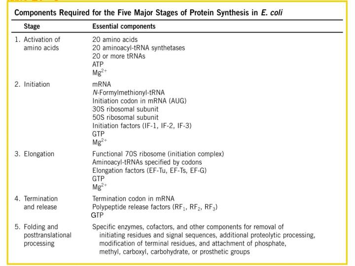
Protein Factors Required for Initiation of Translation in Bacterial Cells
Formation of N-Formylmethionyl-tRNAfMet • A special type of tRNA called tRNAfMet is used here. It is different from tRNAMet that is used for carrying Met to internal AUG codons. The same charging enzyme (synthetase) is believed to be responsible for attaching Met to both tRNA molecules. • Blocking the amino group of Met by a formyl group makes only the carboxyl group available for bonding to another amino acid. Hence, fMet-tRNAfMet is situated only at the N-terminus of a polypeptide chain. • IF2-GTP specifically recognizes fMet-tRNAfMet, which is brought to only the AUG start codon at the P site.
First step in elongation (bacteria): binding of the second aminoacyl-tRNA The second aminoacyl-tRNA enters the A site of the ribosome bound to EF-Tu (shown here as Tu), which also contains GTP. Binding of the second aminoacyl-tRNA to the A site is accompanied by hydrolysis of the GTP to GDP and Pi and release of the EF-Tu•GDP complex from the ribosome. The bound GDP is released when the EF-Tu•GDP complex binds to EF-Ts, and EF-Ts is subsequently released when another molecule of GTP binds to EF-Tu. This recycles EF-Tu and makes it available to repeat the cycle.
Second step in elongation: formation of the first peptide bond The peptidyl transferase catalyzing this reaction is probably the 23S rRNA ribozyme. The N-formylmthionyl group is transferred to the amino group of the second aminoacyl-tRNA in the A site, forming a dipeptidyl-tRNA. At this stage, both tRNAs bound to the ribosome shift position in the 50S subunit to take up a hybrid binding state. The uncharged tRNA shifts so that its 3’ and 5’ ends are in the E site. Similarly, the 3’ and 5’ ends of the peptidyl tRNA shift to the P site. The anticodons remain in the A and P sites.
Third step in elongation: translocation The ribosome moves one codon toward the 3’ end of mRNA, using energy provided by hydrolysis of GTP bound to EF-G (translocase). The dipeptidyl-tRNA is now entirely in the P site, leaving the A site open for the incoming (third) aminoacyl-tRNA. The uncharged tRNA dissociates from the E site, and the elongation cycle begins again.
Termination of protein synthesis in bacteria Termination occurs in response to a termination codon in the A site. First, a release factor (RF1 or RF2 depending on which termination codon is present) binds to the A site. This leads to hydrolysis of the ester linkage between the nascent polypeptide and the tRNA in the P site and release of the completed polypetide. Finally, the mRNA, deacylated tRNA, and release factor leave the ribosome, and the ribosome dissociates into its 30S and 50S subunits.
Energy consumption and rate of translation Energy consumption for synthesis of a polypeptide of N amino acids: N ATPs are required to charge the tRNAs (2N high energy bonds are spent during the charging process). 1 GTP is needed for initiation. N-1 GTPs are required for binding of N-1 aminoacyl-tRNAs to the A site. N-1 GTPs are required for the N-1 translocation steps. 1 GTP is needed during termination. Total: 3N ATPs/GTPs are used. Rate of protein synthesis in E. coli: ~15 aa/second or ~45-nt/second, similar to the elongation speed of RNA polymerase.
Protein synthesis on a circular polyribosome in eukaryotic cells RNA Schematic drawing showing how a series of ribosomes can simultaneously translate the same eukaryotic mRNA molecule, which is in circular form stabilized by interactions between proteins bound at the 3’ and 5’ ends. The 5’ cap and 3’ poly(A) tail have been shown to synergistically enhance translation initiation. They may do so through facilitating ribosome recapture on circularized mRNAs.
Regulation of ferritin mRNA translation Ferritin sequesters iron atoms in the cytoplasm of cells, thereby protecting the cells from the toxic effects of the free metal. Ferritin mRNA translation is controlled by IRP and the intracellular concentration of free iron.
Midterm exam: Monday Nov. 5, 6:30-8:30 p.m. 100 GPB Office hours: Friday, Nov. 3; 3-5 p.m. and Monday, Nov. 5; 2:00-4:00 p.m. Please take a moment to fill out teaching evaluation forms. Thank you for attending the lectures!!!