Download
1 / 25
260 likes | 478 Views
Structural observation of the primary isomerization in vision with femtosecond-stimulated Raman. David W. McCamant et al, Science, 2005, 310, 1006-1009. Miyasaka Laboratory Yusuke Satoh. Vision. (Ref. http://www.kiriya-chem.co.jp/q&a/q52.html). Scheme 1. Structure of eye.

E N D
Structural observation of the primary isomerization in vision with femtosecond-stimulated Raman David W. McCamant et al, Science, 2005, 310, 1006-1009 Miyasaka Laboratory Yusuke Satoh
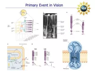
Vision (Ref. http://www.kiriya-chem.co.jp/q&a/q52.html) Scheme 1. Structure of eye The light reaches the retina through eyes and is changed into signal in retina. Signals are sent to our brains.
Retinal Opsin is a protein of 7 spiral structures. A chromophore inside Opsin is Retinal. 11-Cis Retinal changes into all-trans-retinal by light irradiation. Signal is sent to the optic nerve. Scheme 2. Structure of Rhodopsin (Ref. http://www.spring8.or.jp/j/user_info/sp8-info/data/5-6-2k/5-6-2k-3-p394.pdf)
Past research of retinal Table 1 Fluorescence lifetime and Transient absorption spectroscopy of retinal Ref. Chem. Phys. Lett., 2001, 334, 271 Science, 1991, 100, 14526 Transient absorption measurement and time-resolved fluorescence detection of 11-cis Retinal ~200 fs lifetime of the excited state reported.
Motivation But fluorescence and electronic absorption spectra do not provide direct information of the molecular structure. A new time-resolved Raman spectroscopy method is necessary in order to elucidate the dynamics of this isomerization reaction and factors regulating this rapid structural change.
Contents ・Introduction ・Experiment ・Result and Discussion ・Summary
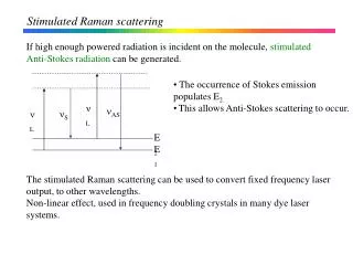
Principle of Spotaneous Raman scattering n0 n0 n0+n n0-n Virtual excited state Ground state n Stokes shift Anti-stokes shift Scheme 3. Mechanism of SpotaneousRaman scattering Raman spectroscopy has been used for the identification of the chemical bond and for the determination of the molecular structure. n0±n: Raman scattering n : Raman shift
Time-resolved Raman spectroscopy Pump pulse Raman scattering n0-n Sample Probe pulse n0 Intermediate Delaytime Detector Scheme. 4 Time-resolved Raman spectroscopy The simple application of femtosecond laser pulse does not provide detailed information of vibrational spectra.
Resonance Raman and Stimulated Raman Excited state Virtual excited state n0 (narrow) n0 n0-n n0-n + (n0-n) (broad) Ground state n n Resonance Raman Stimulated Raman Scheme. 5 Resonance Raman and Stimulated Raman
Stimulated Raman spectroscopy Fig. 1. Stimulated Raman spectroscopy(Ref. Rev. Sci. Instrum., 2004, 75, 4971)
Stimulated Raman system (Ref. Rev. Sci. Instrum., 2004, 75, 4971) Fig. 2 Stimulated Raman spectroscopy system Excited pulse: 500 nm, 30 fs fwhm Raman pump: 805 nm, 3 ps fwhm Raman probe: 830~960 nm, 20 fs fwhm
Structures of 11-cis Retinal and all-trans Retinal Fig. 3 Structure of 11-cis Retinal and all-trans Retinal 11-Cis Retinal change into all-trans Retinal by light irradiation.
Raman spectra of ground-state Retinal ・Raman spectra of 11-cis Retinal(bottom) 1548 cm-1・・・C=C stretch 1100~1300 cm-1・・・C-C single bond stretch and C-H rocking modes 969 cm-1・・・hydrogen-out-of-plane(HOOP) wagging motion of the C11 and C12 hydrogens ・Raman spectra of all-trans Retinal(top) 920, 875, and 850 cm-1・・・C11-H, C10-H, and C12-H wagging mode Fig. 4 Raman spectra of ground-state of 11-cis Retinal(bottom) and all-trans Retinal(top) hydrogen-out-of-plane(HOOP): 水素の面外変角運動 rocking mode:横ゆれ変角運動 wagging mode:縦ゆれ変角運動
Time-resolved Raman spectra of Retinal The dispersive HOOP features evolve on the same time scale as the finger-print bands into the expected three positive features of the Bathorhodopsin spectrum. These data show that there is considerable reactive evolution on the ground-state surface from 200 fs to 1 ps. Fig. 5 Time-resolved Raman spectra of Retinal(200 fs ~1 ps) and Raman spectra of ground state of 11-cis retinal(bottom) and all-trans retinal(top)
Time Profile of C10-H,C11-H and C12-Hhydrogen wagging frequencies Fig. 6 Time profile of C10-H, C11-H and C12-H hydrogen wagging frequency The HOOP frequency increase by 100 cm-1 with 325 fs time constant.
Structures of Retinal, Photorhodopsin and Bathorhodopsin The Bathorhodopsin structure is twisted by –144°about the C11=C12 and by 31°about the C12–C13 bond. The Photorhodopsin structure is more highly distorted, in particular about the C9=C10 (45°), C10–C11 (25°), and C11=C12 (–110°) bonds. With these larger twists, the overall shape of retinal is much more like that of 11-cis Rhodopsin than all-trans Bathorhodopsin, Fig. 7 Retinal chromophore structures for reactant rhodopsin and for photorhodopsin and bathorhodopsin that reproduce the observed hydrogen wagging frequencies.
Theoretical and experimental hydrogen wagging frequencies Caluculated frequency for Photorhodopsin structure show good agreement with experimental data for the C10-H,C11-H modes. Vibrational calculations for the Bathorhodopsin structure yielded features in excellent agreement with experimental data, except for an underestimated C11–H wagging frequency. Fig. 8 Theoretical and experimental hydrogen wagging frequencies for the Photo and Bathorhodopsin structures
The isomerization coordinate for the primary event in vision Excited-state of 11-cis Retinal carry the system toward a conical intersection in ~50 fs. From 200 fs to 1 ps , Photorhdopsin changes into Bathorhodopsin on the ground-state surface. Fig. 9 Multidimensional representation of the isomerization coordinate for the primary event in vision
Summary ・Excited-state decay (200 fs) through a conical intersection is mediated largely by fast HOOP motion. ・By 1 ps, vibrational cooling has narrowed, thereby completing the transformation to Bathorhodopsin.
Stimulated Raman spectroscopy ① Amplitude of coherent vibration induced by Raman and probe pulse Heterodyne detection yields a gain feature on top of the probe envelope in the energy domain shifted in energy relative to the Raman pulse according to the frequency of the vibration. ① Fig. 1 Mechanism of stimulated Raman spectroscopy Stimulated Raman spectroscopy is obtained by this method.
Retinal Opsin is a protein with 7 spiral structures. A chromophore inside Opsin is Retinal. 11-cis retinal changes into all-trans-retinal by light irradiation. Signal is sent to the optic nerve. Scheme 2. Structure of Rhodopsin (Ref. http://www.kiriya-chem.co.jp/q&a/q52.html)
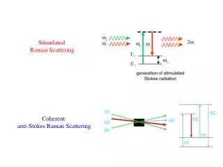
Feynman diagram Feynman diagram
Photoisomerization reaction of Rhodopsin Photoisomerization reaction of Rhodopsin
Principle of Raman scattering (Ref. http://www.natc.co.jp/bunseki/lr.html) Scheme 3. Mechanism of Raman scattering n0±n: Raman scattering n : Raman shift Raman spectroscopy has been used for the identification of the chemical bond and for the determination of the molecular structure.