Download
1 / 36
370 likes | 677 Views
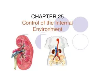
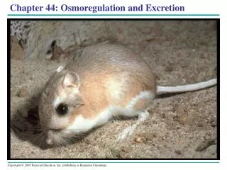
Chapter 44: Osmoregulation and Excretion. A balancing act The physiological systems of animals Operate in a fluid environment The relative concentrations of water and solutes in this environment Must be maintained within narrow limits Homeostasis. Figure 44.1.

E N D
A balancing act • The physiological systems of animals • Operate in a fluid environment • The relative concentrations of water and solutes in this environment • Must be maintained within narrow limits Homeostasis
Figure 44.1 • Freshwater animalsAdaptations for reducing water uptake and conservesolutes • Desert & marine animals desiccating environment, conservewater
Osmoregulation • Balances uptake and loss of water and solutes • based on controlled movement of solutes between internal fluids and the external environment
Osmosis • Osmotic gain and loss of water at the level of the cell • Balanced by osmoregulation
Osmotic Challenges • Osmoconformers • Isoosmotic with their surroundings and do not regulate their osmolarity, e.g. marine inverts. • Osmoregulators expend energy to control water uptake and loss, e.g. marine verts.
Gain of water and salt ions from food and by drinking seawater Osmotic water loss through gills and other parts of body surface Excretion of salt ions from gills Excretion of salt ions and small amounts of water in scanty urine from kidneys • Marine bony fishes are hypoosmotic to sea water • lose water by osmosis and gain salt by both diffusion and from food they eat • Sm. Volume, highly conc. urine Figure 44.3a (a) Osmoregulation in a saltwater fish
Freshwater Animals • Take in water from their hypoosmotic environment • Lose salts by diffusion
Osmotic water gain through gills and other parts of body surface Uptake of water and some ions in food Excretion of large amounts of water in dilute urine from kidneys Uptake of salt ions by gills • Freshwater animals excrete large amounts of diluteurine • Salts lost by diffusion • Are replaced by foods and uptake across the gills Figure 44.3b (b) Osmoregulation in a freshwater fish
Water balance in a human (2,500 mL/day = 100%) Water balance in a kangaroo rat (2 mL/day = 100%) Ingested in food (750) Ingested in food (0.2) Ingested in liquid (1,500) Water gain Derived from metabolism (250) Derived from metabolism (1.8) Feces (0.9) Feces (100) Urine (0.45) Urine (1,500) Water loss Evaporation (900) Evaporation (1.46) Land Animals • Manage their water budget by drinking and by using metabolic water Figure 44.5
Transport Epithelia • Specialized cells that regulate solute movement
Nasal salt gland (a) An albatross’s salt glands empty via a duct into thenostrils, and the salty solution either drips off the tip of the beak or is exhaled in a fine mist. Nostril with salt secretions Lumen of secretory tubule Vein Capillary (c) The secretory cells actively transport salt from theblood into the tubules. Blood flows counter to the flow of salt secretion. By maintaining a concentrationgradient of salt in the tubule (aqua), this countercurrentsystem enhances salt transfer from the blood to the lumen of the tubule. Secretory tubule Artery NaCl Transport epithelium (b) One of several thousand secretory tubules in a salt-excreting gland. Each tubule is lined by a transportepithelium surrounded by capillaries, and drains intoa central duct. Direction of salt movement Blood flow Secretory cell of transport epithelium Central duct • e.g. salt glands of marine birds • remove excess NaCl from the blood Figure 44.7a, b
Proteins Nucleic acids Nitrogenous bases Amino acids –NH2 Amino groups Many reptiles (including birds), insects, land snails Most aquatic animals, including most bony fishes Mammals, most amphibians, sharks, some bony fishes O H C N C HN C O NH2 C C C O N N O NH3 NH2 H H Ammonia Urea Uric acid Nitrogenous Waste • Nitrogenous breakdown products of proteins and nucleic acids Figure 44.8
Ammonia • Animals that excrete ammonia need access to lots of water
Urea • Liver of mammals convert ammonia to less toxic urea • kidneys excreted (w/ minimal loss of water)
Uric Acid • Insects, land snails, reptiles, birds • Little water loss
Capillary Filtration. The excretory tubule collects a filtrate from the blood. Water and solutes are forced by blood pressure across the selectively permeable membranes of a cluster of capillaries and into the excretory tubule. 1 Excretory tubule Filtrate 2 Reabsorption. The transport epithelium reclaims valuable substances from the filtrate and returns them to the body fluids. Secretion. Other substances, such as toxins and excess ions, are extracted from body fluids and added to the contents of the excretory tubule. 3 Excretion. The filtrate leaves the system and the body. 4 Urine Excretory Processes • Urine produced by refining a filtrate derived from body fluids Figure 44.9
Filtration, pressure-filtering of body fluids • Reabsorption, reclaiming valuable solutes • Secretion, addition of toxins and other solutes to the filtrate • Excretion, the filtrate leaves the system
Nucleus of cap cell Cilia Interstitial fluid filters through membrane where cap cell and tubule cell interdigitate (interlock) Tubule cell Flame bulb Protonephridia (tubules) Tubule Nephridiopore in body wall Protonephridia: Flame-Bulb Systems, flatworm Figure 44.10
Coelom Capillary network Bladder Collecting tubule Nephridio- pore Metanephridia Nephrostome Metanephridia, earthworm Figure 44.11
Digestive tract Rectum Hindgut Intestine Malpighian tubules Midgut (stomach) Feces and urine Salt, water, and nitrogenous wastes Anus Malpighian tubule Rectum Reabsorption of H2O, ions, and valuable organic molecules HEMOLYMPH Malpighian Tubules, insects Figure 44.12
Vertebrate Kidneys • Function in both excretion and osmoregulation
Posterior vena cava Renal artery and vein Kidney Aorta Ureter Urinary bladder Urethra (a) Excretory organs and major associated blood vessels Figure 44.13a
Renal medulla Renal cortex Renal pelvis Ureter Section of kidney from a rat Figure 44.13b (b) Kidney structure • 2 distinct regions • An outer renal cortex and an inner renal medulla
Cortical nephron Juxta- medullary nephron Afferent arteriole from renal artery Glomerulus Bowman’s capsule Renal cortex Proximal tubule Peritubularcapillaries Collecting duct SEM 20 µm Distal tubule Efferent arteriole from glomerulus Renal medulla To renal pelvis Collecting duct Branch of renal vein Descending limb Loop of Henle Ascending limb Vasarecta (d) Filtrate and blood flow (c) Nephron Nephron Figure 44.13c, d
Glomerulus of Bowman’s capsule proximal tubule the loop of Henle distal tubule collecting duct
Distal tubule Proximal tubule 4 1 NaCl Nutrients H2O HCO3 H2O K+ NaCl HCO3 NH3 H+ K+ H+ CORTEX Descending limb of loop of Henle 2 Thick segment of ascending limb 3 Filtrate H2O Salts (NaCl and others) HCO3– H+ Urea Glucose; amino acids Some drugs NaCl H2O OUTER MEDULLA NaCl Collecting duct Thin segment of ascending limb 3 5 Urea Key Active transport NaCl H2O Passive transport INNER MEDULLA Filtrate becomes urine Figure 44.14
The mammalian kidney’s ability to conserve water is a key terrestrial adaptation
300 100 300 NaCl H2O Activetransport Osmolarity of interstitial fluid(mosm/L) 400 NaCl H2O Passivetransport 300 NaCl 100 H2O 300 300 CORTEX H2O OUTERMEDULLA NaCl H2O 200 400 400 600 H2O NaCl H2O H2O NaCl H2O H2O 400 600 600 900 H2O NaCl Urea H2O 900 H2O 700 Urea H2O INNERMEDULLA 1200 1200 Urea 1200 • NaCl and urea, contribute to the osmolarity of the interstitial fluid Figure 44.15
Regulation of Kidney Function • Osmolarity of the urine regulated by nervous and hormonal control
Osmoreceptors in hypothalamus Thirst Hypothalamus Drinking reduces blood osmolarity to set point ADH Increased permeability Pituitary gland Distal tubule H2O reab- sorption helps prevent further osmolarity increase STIMULUS: The release of ADH is triggered when osmo- receptor cells in the hypothalamus detect an increase in the osmolarity of the blood Collecting duct Homeostasis: Blood osmolarity • Antidiuretic hormone (ADH) • Increases water reabsorption in the distal tubules and collecting ducts (a) Antidiuretic hormone (ADH) enhances fluid retention by makingthe kidneys reclaim more water. Figure 44.16a
The South American vampire bat, which feeds on blood • Has a unique excretory system in which its kidneys offload much of the water absorbed from a meal by excreting large amounts of dilute urine Figure 44.17
BIRDS AND OTHER REPTILES MAMMALS Bannertail Kangaroo rat (Dipodomys spectabilis) Roadrunner (Geococcyx californianus) Desert iguana (Dipsosaurus dorsalis) Beaver (Castor canadensis) FRESHWATER FISHES AND AMPHIBIANS MARINE BONY FISHES Northern bluefin tuna (Thunnus thynnus) Rainbow trout (Oncorrhynchus mykiss) Frog (Rana temporaria) • Environmental adaptations of the vertebrate kidney Figure 44.18