Download
1 / 24
300 likes | 785 Views
The external beam radiotherapy and Image-guided radiotherapy (1). Somvilai Mayurasakorn , MD. Division of Therapeutic Radiology and Oncology, Faculty of Medicine, Chiang Mai University. Rationale of radiotherapy. MAXIMAL CONTROL OF TUMOR Accuracy of target volume delineation

E N D
The external beam radiotherapy and Image-guided radiotherapy (1) SomvilaiMayurasakorn, MD. Division of Therapeutic Radiology and Oncology, Faculty of Medicine, Chiang Mai University
Rationale of radiotherapy • MAXIMAL CONTROL OF TUMOR • Accuracy of target volume delineation • Precise technique • MININAL NORMAL TISSUE TOXICITY • Sophisticate technique
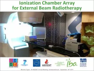
External beam radiotherapy • 2D radiotherapy ICRU 29
External beam radiotherapy • 3D radiotherapy ICRU 29
External beam radiotherapy • Advanced radiotherapy ICRU 62
External beam radiotherapy • Advanced radiotherapy
External beam radiotherapy 2D 3D IMRT Simple to Complex
Advanced radiotherapy treatment Three-dimensional treatment planning systems • Increasingly more conformal treatments • Deliver high dose radiotherapy to an accurately defined target volume • Minimum dose to surrounding tissues • Dose escalation ; leads to improved clinical outcomes • Evidence: lung, head & neck and prostate cance Cancer Imaging (2006) 6, 30–32
Target volumes in EBRT Gross Tumor Volume (GTV) • The gross palpable or visible/demonstrable extent and location of the malignant growth • Supplemental target localization information • CT scan • MRI : improved soft tissue resolution for the definition of soft tissue boundaries, e.g., tumor and surrounding normal tissues • PET: high level of sensitivity and specificity for tumor involvement ICRU IMRT report
Target volumes in EBRT GTV & CTV CT and MRI supplemented by PET information. GTV, light blue; PTV1, yellow; PTV2, red; right parotid gland, dark blue; left parotid gland, orange
Molecular and Functional Imaging Tumor biology plays an important role in • Diagnosis • Treatment decision-making • Assessment of therapeutic response • The development of cellular and molecular imaging • Significant opportunities for the radiation discipline to take the patient’s biological information into the radiation therapy treatment decision-making process and to truly individualize cancer radiotherapy
Molecular and Functional Imaging • Possible to map out the biology distribution • Molecular|functional imaging-guided treatment generally favors non-uniform dose distributions and requires a plan optimization formalism in voxel domain to deal with the biological heterogeneity
Gross Tumor Volume (GTV) Fiberopticexamination Right piriform sinus SCC grade 2 TNM 6thed: T4N0M0 CT MRI T2 FS FDG-PET
Gross Tumor Volume (GTV) PET/CT : target volume of treatment • A prospective trial of PET in RT planning for esophageal carcinoma • significant impact on GTV and PTV • prevented geographic miss • : identifying unsuspected LN involvement Leong T et al.Radiother Oncol 2006;78:254–61
Advanced EBRT • More precious RT • Improves the therapeutic ratio • Reduces normal tissue morbidity • More sophisticate • More resources Adiquate ???
External beam radiotherapy Image-Guided Radiotherapy
SOURCES OF GEOMETRIC UNCERTAINTIES The Netherlands Cancer institute suggested 1. Systematic errors • Uncertainties occurring during treatment preparation • Setup error & organ motion on the planning CT simulator study • Delineation errors • Equipment calibration errors
SOURCES OF GEOMETRIC UNCERTAINTIES The Netherlands Cancer institute suggested 2. Random variations • Uncertainties occurring during treatment execution • Interfraction variations • Intrafraction variations
IGRT • Advanced technique :using imaging devices that allows radiation to be delivered to tumors with more precision than is traditionally possible • Many IGRT technologies are currently available using multiple imaging modalities • USN, video, planar, volumetric
IGRT If we can : • Improve, patient setup (reproducibility) • Reduce uncertainties on patient movements (intra,interfractions) • Reduce uncertainties on target movements (intra,interfractions)
IGRT So we could : • Reduce margins around targets used to take into account movement • Obtain the opportunity to increase dose • Obtain a possible strategy to adaptto target modifications, or to the patient
Imaging techniques for IGRT Video IGRT • Outline of the patient’s features initially seen • As alignment improves, the image becomes less distinct • Finally, a featureless gray image seen