Download
1 / 44
440 likes | 815 Views
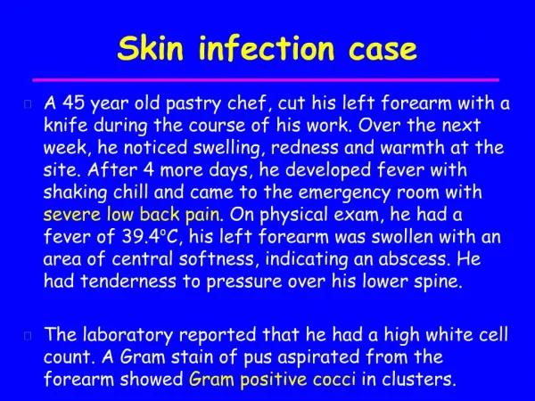
Infection Case Presentation. Dr. Fusun ALATAS Eskisehir Osmangazi University , The Medical School , Department of Chest Diseases. 39 years old , housewife Complaints : Cough , stomach achse , nausea , weakness , dyspnea , sweat History :

E N D
InfectionCasePresentation Dr. Fusun ALATAS Eskisehir Osmangazi University, TheMedicalSchool, Department of ChestDiseases
39 yearsold, housewife Complaints: Cough, stomachachse, nausea, weakness, dyspnea, sweat History: Thepatient had cough, weakness, sweatcompliantsforonemonth, but suddenstartedstomachachse, nauseaanddyspneawereaddedonedayago. Smoking:No Asbest exposure: Environmentalexposurefor 24 years
PhysicalExamination : BP: 110/70 mmHgPulse: 108/min RR:18/minFever: 36.7oC RespiratorySystemExamination: VT andsonoritywasdecreased at rightbasal Crackles at rightmiddlezoneandbreathingsoundsdecreased at basal
Laboratoryfindings: Leucocytes: 10900 mm/3Hb: 13 gr/dlPlt: 135000 mm/3 Sed: 94 mm/h CRP: 27 mg/dl T.protein: 4,1 gr/dl, Alb: 2,2 gr/dl ABG: PaO2: 65 mmHg, PaCO2: 27mmHg O2Sat: %91 pH: 7,51 ECG: Sinustachycardia
Question 1: What is yourdiagnosisforthiscase? A- Lungcancer B- BOOP C- Pulmonarylymphoma D- Pulmonaryembolism E- Parapneumoniceffusion
Question 2: What do you plan fordiagnostictests ? A- BT, thoracentesis B- Ventilation-perfusionscintigraphy, thoracentesis C- Bronchoscopy D- PET-BT E- Dynamic spiral BT, thoracentesis, d-dimerlevels
Thoracentesis: Serousfluid PleuralfluidSerum LDH (U/L) 327 403 Protein (gr/dl) 3.1 4.1 Albumin (gr/dl) 1.8 2.2 Glucose (mg/dl) 72 85 Exudate Slide: PMNL
Question 3: What is yourdiagnosis at this moment? A- Lungcancer + pulmonaryembolism B- Pulmonaryembolism C- BOOP D- Pulmonarylymphoma E- Parapneumoniceffusion + pulmonaryembolism
D- dimer No of CasesSensitivitySpecificity NPV (CI 95%) (CI 95%) (CI 95%) ClassicELISA 1579 97-99 40-46 96-99 Vidas DD 639 98-100 40-49 98-100 ClassicLatex 364 87-96 48-62 83-94 Simplired 1317 82-91 65-71 94-97 Liatest 386 98-100 29-41 96-100
Conditionscauseincreasedlevels of D-dimer • Patients in intensivecareunits • Patients at post-operativeperiod • Peripheralvesseldisease • Cancer • Inflammatorydiseases • Elderlypatients • Trauma Kelly J. ArchInternMed.162;2000
Dynamic spiral BT Diameter of mainpulmonaryartery: app. 24 mm Diameter of rightmainpulmonaryartery: app. 15 mm Diameter of leftmainpulmonaryartery: app. 12 mm Fillingdefectcompatiblewithemboluswasobserved in upperlobeposterordistalbranch of rightlung
Riskfactors • Acquired: • Age • Prolongedimmobilization / trauma / surgery • Cancer • Previous VTE • Comorbidities /obesity • Pregnancy/ Postpartum • Oralcontraceptive • Central venouscathater • Antiphospholipid antibodies • Hyperhomocysteinemia • Longtravels • Hereditary: • FactorV Leiden • ProtrombinG20210 • Deficiency of protein C • Deficiency of protein S • Deficiency of antithrombin III
SLE + APS+ steroidusagewerepresent in patient’shistory. Congenital risk factorwas not determined. Thetreatment of patientwaschangedto LMWH duetocoumadinusagehistory. Increaseddyspnea, purulentsputumandfever (38oC) wereobserved at thirdday of hospitalization.
Sputum; Gram stain: Increased PNL, ARB: (- ) Gram stain: Few gram positivecoccusand gram negativebacillus ARB: (-) Gram stain: Few gram positivecoccusand gram negativebacillus ARB: (-) Sputumculture: negative Bloodculture: negative Pleuralfluidculture: negative
Question 4: Ifthepatientwasdiagnosed as PTE andacceptedhavedepressedimmunity, whatwouldyournext plan? A- Begin3rd generationnonpseudomonalcephalosporintreatment, clinicalandradiologicalfollow-up • B- Begin 2nd generationcephalosporin,performtransthoracicbiopsy • C- Begincarbapenem, follow-uppleuralfluidwithserialthoracentesis • D- Performfiberopticbronchoscopytoevaluateintrabrochialareaandtake sterile samplethenbegincarbapenem+amicasin. Plan transthoracicbiopsy at thesame time. • E- Evaluatethepleurawiththoracoscopyandsamplingifrequired, insert thetubeandbegin 3rd generationnonpseudomonalcephalosporintreatment
FOB findings: Anypathologicalchangeswere not observedexceptextensivepurulentsecretion in tracheaandrightbronchus. Carbapenemtreatmentwasstarted. Sterile BAL: Gram stain: Gram positivecoccus, rare gram negativebacillus ARB: (-)
Sterile BAL culture : Nocardiaspp TMP-SMT wasaddedtotreatment. Carbapenemandamikacinwerestopped at 10thday of treatment.
Pretreatment 20 thday of treatment
Pretreatment 20 thday of treatment
Pretreatment 20 thday of treatment
Pretreatment 20 thday of treatment
PulmonaryNocardiosis • Bacterias of Nocardiaspeciesarepresent in nature, soilandwaterwidespread • Mostlysaprophyte • EdmondNocardwasfirstdescribed in animals in 1888 • Firstdescribed in humansbyEppinger in 1890 Ambrosioni J. Infection 2010; 20
Risk Factors Decreasedcellularimmunity has an important role Transplantationpatients Patientswithleukaemia PatientswithAIDS Patientswhoreceiveprolongedtreatmentswithcorticosteroidsorcytotoxictherapywereunder risk. Cases in normal hostwerealsoreported. Clark NM.Am J Transplant. 2009;9 Suppl 4:S70-7 Martinez R. CurrOpinPulmMed 2008; 14: 219-27.
Thepresentation of pulmonarynocardiosis is highlyvariablebothclinicallyandradiographically. Acute, subacuteandchronicinfectionswithNocardiaspp. havebeenreportedtocause a variety of nonspecificmanifestations, such as anorexia, cough, pleuralpain, dyspnoeaandhaemoptysis. Theincidencewaslowduetomimicedbymalignantandgranulomatousdiseases Conant EF.J ThoracImaging. 1992 ; 7:75-84. Ambrosioni J. Infections 2010; 20
Chestradiographsdemonstrate a variety of findings, such as lobarinfiltrates, cavitation, nodules/massandpleuralinvolvement. Buckley JA. J ComputAssistTomogr 1995; 19: 726-32
Specimens • Bronchialwashing • • BAL • • Sputum • • Abscesses • • Wounddrainages • Tissues • Cerebrospinalfluids Brown –Elliot BA, et al. ClinMicrobiolRev 2006; 19: 259-82.
Theorganism is weaklyacid-fast, but this is usuallylost on subculture. A modifiyeZiehl-Neelsenstain is bestfordemonstratingnocardia. Growth of theorganism in culturemaytake 2-4 weeks Theusage of moleculartechniqueslike PCR arerestricted at the moment
Treatment Therapy is recommendedfor a minimum of 6 weeks, althoughrelapsesoccurlessfrequentlyiftreatment is continuedto 12 weeks. TMP-SMZ (TMP first 4 week 15mg/kg/d, 5 month 10 mg/kg/d) Imipenem4 x 500 mg + Amikacin 1 gr/d Ceftriakson 2 gr/d + Amikacin 1 gr/d Linezolid 2 x 600 mg po • Ambrosioni J. Infections 2010; 20 • Türk Toraks Dergisi 2009; Cilt 10. Ek 5. Vol 10: Suppl 5.
Prognosis The mortality rates were 41% for PN and 64% for disseminated nocardiosis; when Nocardia disseminated to the central nervous system, the mortality was 100%. Martinez R. Respirology 2007;12: 394-400
Masslikeconsolidation Pulmonaryactinomycosis Pulmonarynocardiosis • Pulmonarylymphoma Pulmonaryaspergillosis BAC Lipoidpneumonia