Download
1 / 179
1.84k likes | 2.78k Views
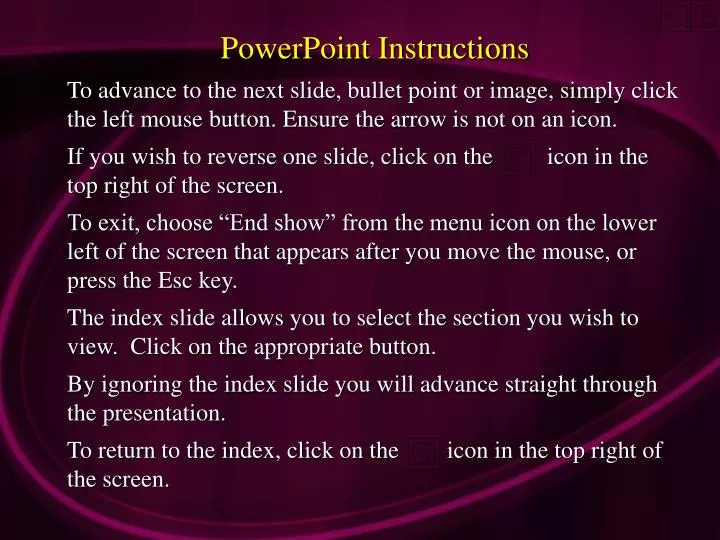
PowerPoint Instructions To advance to the next slide, bullet point or image, simply click the left mouse button. Ensure the arrow is not on an icon. If you wish to reverse one slide, click on the icon in the top right of the screen.

E N D
PowerPoint Instructions To advance to the next slide, bullet point or image, simply click the left mouse button. Ensure the arrow is not on an icon. If you wish to reverse one slide, click on the icon in the top right of the screen. To exit, choose “End show” from the menu icon on the lower left of the screen that appears after you move the mouse, or press the Esc key. The index slide allows you to select the section you wish to view. Click on the appropriate button. By ignoring the index slide you will advance straight through the presentation. To return to the index, click on the icon in the top right of the screen.
Caries Diagnosis and How to Use the DIAGNOdent E-mail: gwmilicich@xtra.co.nz Website: www.advancedental-ltd.com
INDEX To exit. Press Esc. Traditional caries diagnostic model Current caries model Caries detection dye (CDD) How to use the DIAGNOdent Interpreting the results False positives Hypomineralized (hypocalcific) enamel Hidden caries Variable readings E-mail: gwmilicich@xtra.co.nz Website: www.advancedental-ltd.com Fissure sealants
Traditional DiagnosticModel Low sensitivity High specificity
Low Sensitivity Conventional diagnosis can miss significant amounts of decay High Specificity Conventional diagnosis does not produce a lot of false positive diagnoses
Traditional fissure caries model Probe does not stick “No caries”
Traditional fissure caries model Probe will now stick Enamel decalcification
Traditional fissure caries model Continuing decalcification finally leads to cavitation of the enamel
Traditional fissure caries model Continuing decalcification finally leads to cavitation of the enamel
Traditional fissure caries model Continuing acid attack leads to dentin caries and further cavitation
Penning C, van Amerongen JP, Seef RE & ten Cate JM.Validity of probing, for fissure caries diagnosis.Caries Res 26(6):445-9, 1993 “ Probing found unreliable in finding fissure caries”
Black, G.V. Operative Dentistry. Vol. I Henry Kimpton, London. 7th Ed, p32, 1924 “ A sharp explorer should be used with some pressure and if a very slight pull is required to remove it, the pit should be marked for restoration even if there are no signs of decay.”
ROCK WP, KIDD EAM.Br Dent J. 164(8): 243-47, 1988. “… decay is difficult to detect in radiographs unless larger than 2mm to 3mm deep into dentin, or 1/3 the bucco-lingual distance.”
4mm deep, total decalcification.Cavity was widened to 1/3 occlusal width to show on Xray 4mm 1/3 occlusal width
X-RAY Contact point caries is much easier to detect radiographically 1/3rd Digitally created
Chan DCN. Current methods and criteria for finding decay in North America.J Dent Ed 57(6):422-425, 1993 Caries is regularly found beneath a seemingly intact enamel surface Frequently the diagnosis of occlusal caries is less than straightforward
AL-SEHAIBANY, WHITE & RAINEYJ Clin Pediatr Dent 20(4):293-298 1996 The reliability of carious lesion diagnosis by sharp explorer compared to diagnosis of carious lesion by histological cross section was 25%. ______________________________ A seemingly intact occlusal enamel surface may conceal an extensive lesion of the dentin
We are just guessing... when we apply the current standards of care in 21st century operative dentistry
The traditional fissure caries diagnosticmodel is very crudeLOW SENSITIVITY
Modern fissure caries model Organic plug
Modern fissure caries model Decalcified or hypocalcific enamel Organic plug Acid percolation through porous, hypocalcific enamel can lead to failure of the organic plug
Modern fissure caries model Enamel may be developmentally hypomineralized, and consequently porous through its full thickness Consequently, dentin can be exposed to acid without cavitation of the enamel leading to developmental dentin caries ACID
Modern fissure caries model OR by the time the tooth has emerged from under the operculum, the fissure enamel can already be carious ACID
Modern fissure caries model These areas may not be decalcified, and a probe won’t stick
Modern fissure caries model Fissure walls are in close apposition Once the organic plug fails, bacteria have access to the depths of the fissure A probe will be unable to detect caries here Decalcification
Modern fissure caries model Presentation is inverted compared to the traditional model Continuing decalcification +dentin caries
Modern fissure caries model Can’t diagnose this with a probe or Caries Detection Dye (CDD) Defects in the fissure walls can lead to dentin caries with NO enamel decalcification
Modern Fissure CariesAnatomy Model(Summary of realistic ‘coke bottle ‘ shape) Organic plug Decalcified or hypocalcific enamel (caries in this zone is undetectable by probe) (This area may not be decalcified thus a probe won’t stick) Enamel defects in fissure wall De-mineralizing dentin
Fissure Caries • The DIAGNOdent can “read” 2mm into the tooth • As long as the fissure is cleaned of debris, readings will detect changes in the underlying enamel and dentin • The use of caries detection dye (CDD) to stain porous, carious enamel will help identify carious tooth structure that needs removing
How Does Caries Detection Dye Work Fusayama T. A Simple Pain –Free Adhesive Restorative System.1:18 1993 “The mechanism of differential staining does not involve selective chemical bonding of the dye in usual staining, but the selective penetration of the solvent”
How Does Caries Detection Dye Work Fusayama T. A Simple Pain –Free Adhesive Restorative System.1:18 1993 It is simply filling the voids in enamel and dentin that are created by acid attack, or filling voids present in hypomineralized enamel
Slow onset caries Caries Detection Dye SEM Haikel et al.1983 Enamel prisms remain, but with some mineral loss P Loss of interprismatic enamel creates a “micro-pore” effect S
AL-SEHAIBANY F, WHITE G & RAINEY J.T.J Clin Pediatr Dent 20(4):293-298, 1996 CDD is a reliable diagnostic tool for occlusal carious lesions. Ratio of occlusal grooves stained by dye, to underlying carious lesions, is 1:1 by histological x-section in extracted teeth 75% of occlusal carious lesions missed by probing were found using CDD
Carious fissure walls in very close apposition Fissure appears totally sound
Carious fissure walls in very close apposition Fissure appears totally sound Carious (decalcified) enamel in the depths of the fissure Stained with Caries Detection Dye
CDD Carious fissure walls in very close apposition
Stained with CDD This tooth was partially erupted under an operculum for 18 months. CDD has stained the carious enamel.
Carious enamel and dentin stained Note diffusion of the dye into demineralized occlusal enamel, as well as into the fissure