Download
1 / 48
500 likes | 1.15k Views
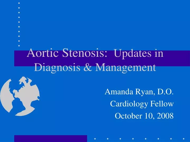
Aortic Stenosis: Updates in Diagnosis & Management. Amanda Ryan, D.O. Cardiology Fellow October 10, 2008. Learning Objectives. Following the presentation the participant should be able to: Define aortic stenosis based on echocardiographic guidelines Explain primary causes and risk factors

E N D
Aortic Stenosis: Updates in Diagnosis & Management Amanda Ryan, D.O. Cardiology Fellow October 10, 2008
Learning Objectives • Following the presentation the participant should be able to: • Define aortic stenosis based on echocardiographic guidelines • Explain primary causes and risk factors • Identify the demographics • Discuss optimal management including timing of valvular surgery
Disclosure of commercial support • With respect to my presentation, I DO NOT have any financial arrangements or affiliation with any corporate organization associated with the manufacture, distribution, or promotion of a drug or device which is related to the topic of my presentation.
Aortic Valve Aortic Valve
Aortic Stenosis Definition • Disease in which progressive obstruction to LV outflow results in: • pressure hypertrophy of the LV • symptoms of angina, dyspnea, and syncope (classic triad) • if untreated, will lead to death
Causes of Aortic Stenosis • Supravalvular • Subvalvular • discrete • tunnel • Valvular • congenital (1-30yrs old) • bicuspid (40-60yrs old) • rheumatic (40-60yrs old) • senile degenerative (>70yrs old)
This is the aortic valve viewed from the outflow side, i.e. from the aorta. Blood flows up through the valve from the left ventricle, pushing the cusps out of the way. Two of the three cusps are clearly visible in this view.
Schematic of AV Tricuspid (left) and Bicuspid (right) Aortic Valve.
Supravalvular • congenital abnormality in which ascending aorta superior to the aortic valve is narrowed • rarest site of AS • either a single discrete constriction or a long tubular narrowing
Supravalvular cont • Dx should be suspected in young pt with LVOT murmur • On physical exam - thrill felt on palpation of right carotid but not left • On 2D echo - visualization of narrowed ascending aorta • On Doppler - provides info on magnitude of obstruction
Suprvalvular cont • Associations: • Elfin facies • Hypercalcemia • Peripheral pulmonic stenosis
Subvalvular AS • Discrete seen in 10% of all pts with AS • can be secondary to a subvalvular ridge that extends into LVOT or to a tunnel-like narrowing of the outflow tract • Aortic regurgitation frequently accompanies
Subvalvular cont • Echo - visualization of a narrowing or discrete subvalvular ridge extending into the LVOT and a high-velocity turbulence on continuous wave doppler • If site of obstruction is not visualized on transthoracic echo, TEE is indicated
Subvalvular vs HCM • Dx of subvalvular AS needs to be differentiated from dynamic outflow obstruction of HCM b/c tx differs • Discrete subvalvular - some recommend resection in all pts with moderate or higher to relieve degree of LVOT obstruction and prevent progressive AR
Valvular • Accounts for most cases • Cause of valve abnormality depends on age at presentation • Teens to early 20’s - congenitally unicuspid or fused bicuspid valve • 40’s to 60’s - calcified bicuspid or rheumatic disease • 70’s and beyond - senile degeneration of valve with calcific deposits
Pathophysiology • In adults with AS, obstruction develops gradually, usually over years • LV adapts to systolic pressure overload through a hypertrophic process that results in increased LV wall thickness (normal chamber volume maintained) • Eventually, LV cannot compensate for the long-standing pressure overload and ventricular dilation and progressive decrease in systolic function
Pathophysiology 1. increase in afterload 2. decrease in systemic & coronary blood flow from obstruction 3. progressive hypertrophy
Pathophysiology • Depressed contractile state of the myocardium may also cause low EF • Difficult to determine whether low EF is secondary to this or to excessive afterload • When caused by depressed contractility, corrective surgery is less beneficial.
More Pathophysiology • Exertional dyspnea is common, even when LVSF is preserved • Diastolic dysfunction is common and result in increased LV filling pressures that are reflected onto pulmonary circulation • Diastolic dysfunction occurs from prolonged ventricular relaxation and decreased compliance and is caused by myocardial ischemia, a thick non-compliant ventricle, and increased afterload
Angina • Exertional angina may occur in absence of epicardial coronary artery obstruction • Pathophysiology here is related to mismatch of myocardial oxygen supply and demand due to • high diastolic pressures • decreased myocardial perfusion gradient • increased myocardial mass
Critical AS • In critical AS, with onset of systemic hypotension (either from medications or vasovagal reaction), coronary artery perfusion may decrease. • This increased myocardial oxygen supply/demand mismatch and results in myocardial ischemia • This reduces forward cardiac output, aortic diastolic pressure decreases, which further decreases coronary perfusion.
Clinical Presentation • Asymptomatic but heart murmur detected on physical exam • May have any one or all three exertional dyspnea, angina, syncope • Occasionally pt with end stage aortic stenosis and LV dysfunction present with anasarca and cardiac cachexia
Coagulation Abnormalities • In most pts with severe AS, impaired platelet fxn and decreased levels of von Willebrand factor are noted • Severity of coagulation problem correlates with degree of AS • Associated with clinical bleeding in 20% of patients • Resolves after valve replacement
Physical Exam • Dampened upstroke of carotid artery • Sustained bifid LV impulse • Single or split S2 • Late peaking systolic ejection murmur (may be heard with same intensity at apex and base) • The severity more related with timing of peak and duration than loudness
EKG and Chest X-ray • EKG usually shows NSR with LVH • if afib is present, concomitant mitral valve disease or thyroid disease should be suspected • Chest radiography may show LV predominance with dilatation of ascending aorta. Calcification of aorta often seen on lateral films
Echocardiography • 2D and Doppler echo are imaging modalities of choice to diagnose and estimate severity of AS • 2D can identify location • In pts with valvular, the cause may be assessed in parasternal short-axis view • Doppler excellent for assessment of severity
Class 1 Echo recommendations • Echocardiography is recommended for diagnosis and severity of AS • Echocardiography is recommended in patients with AS for assessment of LV wall thickness, size, and function • Echocardiography is recommended in patients with known AS and changing symptoms • Echocardiography is recommended for assessment of changes in hemodynamic severity and LV function in pts with known AS during pregnancy • Transthoracic echocardiography is recommended for re-evaluation of asymptomatic patients: severe AS - yearly; moderate AS - every 1-2 years; mild AS - every 3-5 years
Doppler • Modified Bernoulli equation (delta P=4v2), a maximal instantaneous and mean aortic valve gradient can be derived from continous pulse wave doppler velocity across aortic valve. • The accuracy of the above relies on the fact that Doppler beam is parallel to the stenotic jet
More Doppler Data • Aortic valve gradients depend on severity of obstruction and on flow. • Pt may have low cardiac output and gradient less than 40mm Hg, but still have severe stenosis. • Aortic valve area (AVA) is used to overcome this limitation
Class 1 Indications for Cardiac Catheterization • Coronary angiography is recommended before AVR in pts with AS at risk for CAD • Cardiac cath for hemodynamic measurements is recommended for assessment of severity of AS in symptomatic pts when noninvasive tests are inconclusive or there is a discrepancy between non-invasive tests and clinical findings • Coronary angiography is recommended before AVR in pts with AS for whom a pulmonary autograft (Ross procedure) is contemplated and if the origin of the coronary arteries is not identified by noninvasive techniques
Aortic Valve Area • In cardiac cath lab, AVA is calcuated from pressure gradient and an independent measure of cardiac output • AVA = 1000 X CO 44 X SEP X HR X s.r of delta P SEP is systolic ejection period P is pressure difference across valve
AVA cont • Echo and doppler estimate aortic valve area by the continuity equation AVA = LVOTarea X LVOTtvi AVtvi AV = aortic valve flow velocity TVI = time-velocity integral • Doppler echo may underestimate AV gradient
Natural History • After symptoms occur in a pt with severe AS, rapidly progressive downhill course • 2 to 3 year mortality of 50% • Therefore, recommendations support AV replacement in all pts with severe AS and symptoms • In young, healthy pts, very low perioperative mortality of 1-2%
Asymptomatic AS • Controversial recommendations regarding valve replacement • Some studies have shown increased mortality in asymptomatic pts while others have shown similar mortality to age-matched normal adults • Frequent reassessment for symptoms
Calcific Aortic StenosisNote that there are three distinct cusps in this valve, and that the free edges of the cusps appear normal. Heaped up calcific deposits extend from the cusps into the sinuses of Valsalva. This calcification prevents normal opening of the valve. Calcification of the aortic valve may be a result of rheumatic heart disease. The gross appearance illustrated here, however, is characteristic of degenerative ("wear and tear") calcific aortic stenosis in an elderly individual. Bicuspid aortic valves (a congenital abnormality) also tend to calcify, but usually at an earlier age than normal three-cusped valves.
CT Dx of AS CT Evaluation of Aortic Stenosis • Excellent Assessment of Calcification • May establish stenosis severity • Cannot establish insufficiency • Good evaluation of mechanical prostheses • Evaluates coronary anatomy in patients undergoing valve surgery
This enhanced CT image puts you inside the aorta, looking down at the aortic valve. You can clearly see the three valve leaflets, currently closed.
Cardiac MRI & AS CMR Evaluation of Aortic Stenosis • Safe • Minimally invasive • Absence of ionizing radiation • Absence of nephrotoxic contrast agents • Morphology + physiology • Simultaneous cardiac evaluation
Medical Therapy • Antibiotic prophylaxis is NOT recommended in all pts with AS for prevention of infective endocarditis. • Pts with associated systemic HTN should be treated cautiously with appropriate antihypertensives (preload dependence) • Statins have been studied to see if they cause regression or delayed progression of leaflet calcification (need more data)
Class 1 Recommendations for Aortic Valve Replacement in AS • AVR is indicated for symptomatic pts with severe AS • AVR is indicated for pts with severe AS undergoing CABG • AVR is indicated for pts with severe AS undergoing surgery on aorta or other heart valves • AVR is recommended for pts with severe AS and LV systolic dysfunction (EF<50%)
Aortic Valve Surgery • Options include: • AVR with mechanical or bioprosthetic valve • AVR with allograft (homograft) • Pulmonic vavle autotransplantation (Ross) • Aortic valve repair • LV to descending aorta shunt
Types of AVR Examples of replacement aortic valves: a) shows an aortic homograft, b) and c) show a xenograft, d) shows a ball and cage valve, e) shows a tilting-disk valve, f) shows a bi-leaflet valve
Elderly Patients • Pts >80years, operative mortality as high as 30%. • Percutaneous aortic balloon valvuloplasty is an alternative to valve replacement introduced in ‘80s. • Inflating one or more large balloons across the aortic valve from a percutaneous route, a modest decrease in gradient and improvement in symptoms
Balloon Valvuloplasty • Follow-up has demonstrated a high rate of re-stenosis (>60% at 6 months and nearly 100% at 2 years), with no decrease in mortality rate after procedure • Therefore, now only has a role in critically ill elderly pts who are not candidates for surgery or as a “bridge” in critically ill pts before AV replacement
References • Bonow RO, Carabello BA, Chatterjee K, de Leon AC Jr., Faxon DP, Freed MD, Gaasch WH, Lytle BW, Nishimura RA, O’Gara PT, O’Rourke RA, Otto CM, Shah PM, Shanewise JS. ACC/AHA 2006 guidelines for the management of patients with valvular heart disease: a report of the American College of Cardiology/America Heart Association Task Force on Practice Guidelines (Writing Committee to Develop Guidelines for the Management of Patients With Valvula Heart Disease). American Heart Association Web Site. Available at: http://www.americanheart.org. Circulation. • Rosenhek R, Klaar U, Schemper M, et al. Mild and moderate aortic stenosis. Natural history and risk stratification by echocardiography. Eur Heart J 2004;25:199 –205. • Cosmi JE, Kort S, Tunick PA, et al. The risk of the development of aortic stenosis in patients with “benign” aortic valve thickening. Arch Intern Med 2002;162:2345–7. • Stewart BF, Siscovick D, Lind BK, et al. Clinical factors associate with calcific aortic valve disease: Cardiovascular Health Study. J Am Coll Cardiol 1997;29:630–4.