Download
1 / 27
400 likes | 1.78k Views
Brachial Plexus & Lumbosacral Plexus. Dr. Saeed Vohra & Dr.Sanaa Alshaarawy. Objectives. At the end of this lecture, the students should be able to : Describe the formation of brachial plexus (site, roots) List the main branches of brachial plexus

E N D
Brachial Plexus & Lumbosacral Plexus Dr. Saeed Vohra & Dr.SanaaAlshaarawy
Objectives • At the end of this lecture, the students should be able to : • Describe the formation of brachial plexus (site, roots) • List the main branches of brachial plexus • Describe the formation of lumbosacral plexus (site, roots) • List the main branches of lumbosacral plexus • Describe the important Applied Anatomy related to the brachial & lumbosacral plexuses
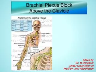
Formation of Brachial Plexuses • It is formed in theposterior triangleof the neck. • It is the union oftheanterior ramiof the 5th ,6th ,7th ,8th cervical and the 1st thoracic spinal nerves
Divisions • The plexus is divided into • Roots • Trunks • Divisions • Cords • Terminal branches
Trunks • Upper trunk • Union of the rootsofC5 & 6 • Middle trunk • Continuation of theroot ofC7 • Lower trunk • Union of the rootsofC8 & T1
Divisions & Cords • Each trunk divides into anterior and posterior division • Posterior cord: • From the three posterior divisions • Lateral cord: • From the anterior divisions of the upper and middle cords
CORDS & BRANCHES • Medial cord • It is the continuation of the anterior division of the lower trunk • Branches • All three cords will give branches, those will supply their respective regions
lateral Cord (2LM) posterior Cord (ULTRA) medial Cord (4MU) The Brachial Plexus Long Thoracic (C5,6,7) Nerve to Subclavius(C5,6) Dorsal Scapular(C5) Suprascapular(C5,6) upper trunk roots C5 C6 middle trunk C7 lower trunk C8 T1 Anterior divisions Posterior divisions
The Plexus can be divided into 5 stages: • Roots:in the posterior∆ of the neck • Trunks: in the posterior∆ of the neck • Divisions: behind the clavicle • Cords:in the axilla • Branches: in the axilla • The first 2 stages lie in the posterior triangle, while the last 2 sages lie in the axilla.
A. Branches from Roots • 1. Nerve torhomboids (dorsal scapular nerve) C5 • 2. Long thoracic nerve C5, 6 & 7 B. Branches from Trunk • Nerve to subclavius • Suprascapularnerve (supplies supraspinatus & infraspinatus)
(C)Branches from Cords Lateral Cord (2LM) .Lateral pectoral n .Lateral root to median n .Musculocutaneous n C5 C6 C7 C8 T1 Medial cord (4MU) .Medial pectoral n. .Medial root to median n. .Medial cutaneous n of arm. .Medial cutaneous n of forearm. .Ulnar n. Posterior Cord (ULTRA) .Upper subscapular n .Lower subscapular n .Thoracodorsal n .Radial n .Axillary n
Brachial Plexus Injuries • Upper Lesions of the Brachial Plexus Upper Trunk C5,6 (Erb-Duchenne Palsy ”waiter's tip position”. • Resulting from excessive displacement of the head to the opposite side and depression of the shoulder on the sameside(a blow or fall on shoulder). • The position of the upper limb in this condition has been likened to that of a porter or waiter hinting for a tip or policeman’s tip hand. • The arm hangs by the side and is rotated medially. The forearm is extended and pronated Erb-Duchenne’s paralysis due to injury of Upper Trunk of Brachial Plexus.
Brachial Plexus Injuries • Lower Lesions of the Brachial Plexus, (Klumpke Palsy)/LowerTrunk(C8,T1)Lesion • Lower lesions of the brachial plexus are usually traction injuries caused by a person falling from a height clutching at an object to save himself. The first thoracic nerve is usually torn. • The nerve fibers from this segment run in the ulnar and median nerves to supply all the small muscles of the hand. The hand has a clawed appearance due to ulnar nerve injury Claw Hand Claw Hand Hand of Benediction or Pop’s Blessings(APE HAND) willresult from median nerve injury.
LUMBAR PLEXUS • Formation: By ventral rami of L1, 2, 3 and most of L4 • Site: In the substance of psoas major muscle • Main branches: Iliohypogastric & ilioinguinal (L1) to anterior abdominal wall Obturator(L2-L4) to medial compartment of thigh Femoral (L2-L4) to anterior compartment of thigh
FEMORAL NERVE • Origin: • A branch from lumbar plexus (L2,3,4) • Course: • Descends lateral to psoas major & enters the thigh behind the inguinal ligament • Passes lateral to femoral artery & divides into anterior & posterior divisions.
FEMORAL NERVE INJURY • Motor effect: • Wasting of quadriceps femoris • Loss of extension of knee • Weak flexion of hip (psoas major is intact) • Sensory effect: • loss of sensation over areas supplied antero-medial aspect of thigh & medial side of leg & foot
SACRAL PLEXUS • Formation: By ventral rami of a part of L4 & whole L5 (lumbosacral trunk) + S1, 2, 3 and most of the S4 • Site: In front of piriformismsucle
SACRAL PLEXUS • Main branches: Pelvic splanchnic nerve preganglionic parasympathetic to pelvic viscera & hindgut Pudendal nerve to perineum Sciatic nerve to lower limb
SCIATIC NERVEThe largest nerve of the body • Origin: • from sacral plexus (L4, 5, S1, 2, & 3) • Course: • Leaves the pelvis through greater sciatic foramen, below piriformis & passes in the gluteal region (between ischial tuberosity & greater trochanter) then to posterior compartment of thigh • Divides into tibial & common peroneal (fibular) nerves Ischial tuberosity
TIBIAL NERVE • Course: • Descends through popliteal fossa to posterior compartment of leg, accompanied with posterior tibial vessels • Passes deep to flexor retinaculum to reach the sole of foot where it divides into 2 terminal branches
COMMON PERONEAL (FIBULAR) NERVE • Course: • Leaves popliteal fossa & turns around the lateral aspect of neck of fibula Then divides into: • Superficial peroneal: descends into lateral compartment of leg • Deep peroneal: descends into anterior compartment of leg
SUMMARY • The lumbar plexus is formed by ventral rami of L1,2,3 and most of L4, in substance of psoas major muscle • The sacral plexus is formed by ventral rami of a part of L4 & whole L5 (lumbosacral trunk) plus the S1,2,3 and most of S4, in front of piriformismsucle. • The femoral nerve, a branch of lumbar plexus (L2,3,4). • Its injury will affect the flexion of hip & extension of knee as well as loss of sensation of skin of anteromedial aspects of the thigh, medial side of knee, leg and foot • The sciatic nerve is a branch of sacral plexus (L4,5, S1,2,3) • Its injury will affect the flexion of knee, extension of hip, all movements of leg & foot, as well as loss of sensation of skin of leg & foot (except areas supplied by saphenous branch of femoral nerve)
1. Lesion of the upper trunk of the brachial plexus leads to : • Klumpke palsy. • Erb-Duchenne palsy • Drop wrist & hand. • Ape hand. • 2. Which one of the following nerves is a branch of posterior cord of brachial plexus? • Ulnar • Radial • Median • Musclocutanous
QUESTION 1 • The femoral nerve supplies: • Extensors of hip. • Skin of dorsum of foot. • Hamstrings. • Extensors of knee
QUESTION 2 • Injury of common peroneal nerve leads to: • Loss of dorsiflexion of ankle • Loss of inversion of foot • Loss of extension of knee • Loss of flexion of toes