Download
1 / 18
180 likes | 330 Views
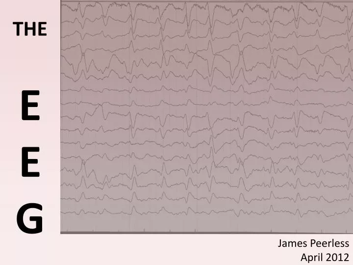
E E G. THE. James Peerless April 2012. Objectives. Physics and Clinical Measurement Anaesthesia for neurosurgery, neuroradiology and neurocritical care Demonstrates knowledge of: PC_BK_52: Amplification of biological signals: including ECG, EMG, EEG, BIS, CFM, CFAM

E N D
E EG THE James Peerless April 2012
Objectives Physics and Clinical Measurement Anaesthesia for neurosurgery, neuroradiology and neurocritical care Demonstrates knowledge of: • PC_BK_52: Amplification of biological signals: including ECG, EMG, EEG, BIS, CFM, CFAM • NA_IK_04: Explains the indications for using neurophysiological monitoring [including EEG, evoked potentials and ICP measurement] to benefit patients requiring neurosurgery/neuro-critical care
History • 1875 – electrical activity from animals’ brains • 1890 – electrical activity altered by stimuli • 1924 – first human EEG described • 1934 – epileptiform activity demonstrated
Introduction • Recording of electrical activity of the brain • Signals from ~20 scalp electrodes are collated and presented as 16 traces • 10-30 minutes; recorded with video to correlate brain activity with clinical picture • Characteristics of the traces, i.e. shape, distribution, incidence and symmetry are analysed
What is it? • There are millions of nervous action potentials firing at any one time • ‘Brain waves’ are the summation of synchronous activity of neurons detected at the scalp • Brain activity shows oscillation at various frequencies
Method • Electrodes • Amplifier • Filter • Microprocessor • Output Monitor
Biological Signal Transduction • Heart – ECG • 0.05 – 100 Hz • 1mV • Brain – EEG • 0 – 13 Hz • 50 – 200 μV • Muscle – EMG • 1 – 20 000 Hz • 1 mV
Current Uses in Medicine • Clinical medicine • Distinguishing between seizure types • Monitoring of depth of anaesthesia • BIS • indicator of cerebral perfusion in carotid endarterectomy • Intensive & Neurocritical care • brain function monitoring • to monitor for non-convulsive seizures/ status epilepticus • to monitor levels of sedation • Research
Anaesthesia & The EEG • Why don’t we use it much? • Expensive equipment • Skilled operators • Dissimilar anaesthetic agents generate different EEG patterns or signatures • Increasing depth of anaesthesia signal amplitude is decreased, frequency increases
Causes of EEG Depression • EEGs change with age, state of consciousness (incl. GA) • Metabolic states (e.g. hypoglycaemia, hepatic coma) • Hypotension, hypoxia, hypercarbia, cerebral oedema • Encephalitis • CJD • Brain death isoelectric (flat line)
BIS • Bispectral index analysis • Monitors electrical activity and quantifies level of sedation • Aims: to reduce awareness; reduce over-/underdosing of drugs • Works best with hypnotic agents • Doesn’t work with ketamine; and less sensitive to sedative effect of opioids
BIS • Displayed as a continuous trend • Facial electromyogram (EMG) • BIS • Signal Quality Index (SQI) • Forehead sensor • 4 tines
Summary • EEG measures electrical activity from the brain • Complex analysis limits its use in mainstream anaesthetic practice • BIS monitoring incorporates EEG and quantifies depth of anaesthesia
MCQ Concerning electroencephalography (EEG): • Voltages are in the range of 10-100 millivolts • Spontaneous EEG activity is lost when the body temperature drops below 25 °C • β waves are enhanced by sedatives • δ waves only occur in brain injury • θ waves occur at a frequency of 4-7 Hz
MCQ Concerning electroencephalography (EEG): • Voltages are in the range of 10-100 millivolts • Spontaneous EEG activity is lost when the body temperature drops below 25 °C • β waves are enhanced by sedatives • δ waves only occur in brain injury • θ waves occur at a frequency of 4-7 Hz
MCQ Regarding the BIS monitor: • It uses a dimensionless scale from 0 to 100 Hz • Hypothermia can increase the BIS value • The BIS value is not accurate during ketamine anaesthesia • Interference can occur due to EMG or diathermy • BIS can measure the concentration of a particular drug
MCQ Regarding the BIS monitor: • It uses a dimensionless scale from 0 to 100 Hz • Hypothermia can increase the BIS value • The BIS value is not accurate during ketamine anaesthesia • Interference can occur due to EMG or diathermy • BIS can measure the concentration of a particular drug