Download
1 / 1
10 likes | 260 Views
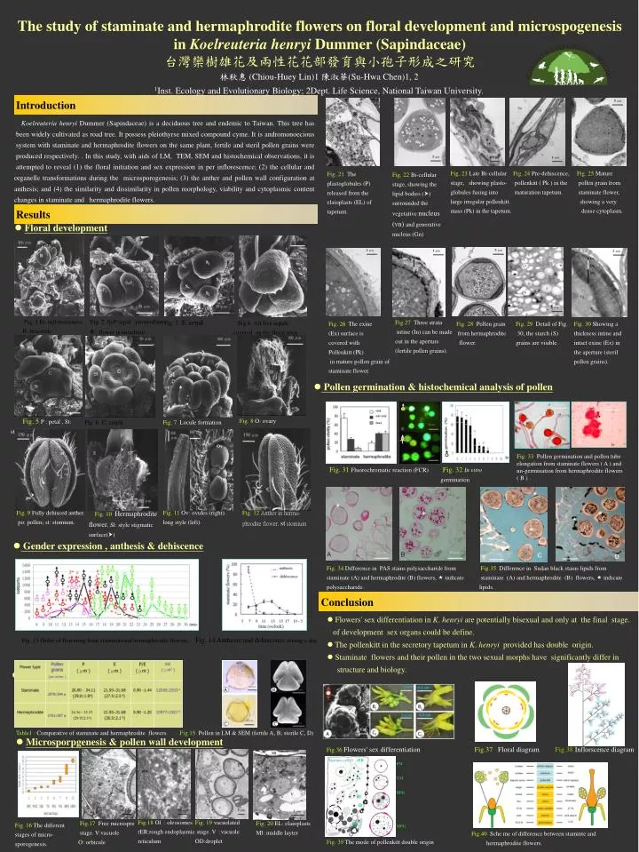
The study of staminate and hermaphrodite flowers on floral development and microspogenesis in Koelreuteria henryi Dummer (Sapindaceae) 台灣欒樹雄花及兩性花花部發育與小孢子形成之研究. rER. Tapetum cell25. FM VM BPG MPG. 林秋惠 (Chiou-Huey Lin)1 陳淑華 (Su-Hwa Chen)1, 2

E N D
The study ofstaminate and hermaphrodite flowers on floral development and microspogenesis in Koelreuteria henryi Dummer (Sapindaceae) 台灣欒樹雄花及兩性花花部發育與小孢子形成之研究 rER Tapetum cell25 FM VM BPG MPG 林秋惠 (Chiou-Huey Lin)1 陳淑華(Su-Hwa Chen)1, 2 1Inst. Ecology and Evolutionary Biology; 2Dept. Life Science, National Taiwan University. Ol El O PK FM • Fig. 23Late Bi-cellular • stage, showing plasto-globules fusing into • large irregular pollenkitt. • mass (Pk) in the tapetum. • Fig. 24Pre-dehiscence, • pollenkitt ( Pk ) in the • maturation tapetum. • Fig. 25Mature • pollen grain from • staminate flower, • showing a very • dense cytoplasm. • Fig. 21The plastoglobules (P) released from the elaioplasts (EL) of tapetum. • Fig. 22Bi-cellular stage, showing the lipid bodies () surrounded the vegetative nucleus(vn)and generative nucleus (Gn) Ex • Fig. 1 In: inflorescences B :bracteole Fig. 2 SeP:sepal primordium : flower primordium • Fig. 3 S: sepal • Fig.27 Three strata • intine (In) can be made out in the aperture (fertile pollen grains). • Fig.4All five sepals covered on the floral apex. • Fig. 26The exine • (Ex) surface is • covered with • Pollenkitt (Pk) • in mature pollen grain of staminate flower. • Fig. 28Pollen grain • from hermaphrodite • flower. • Fig. 29Detail of Fig. • 30, the starch (S) grains are visible. • Fig. 30Showing a thickness intine and intact exine (Ex) in the aperture (steril pollen grains). • Pollen germination & histochemical analysis of pollen • Fig. 33Pollen germination and pollen tube • elongation from staminate flowers ( A ) and • un-germination from hermaphrodite flowers • ( B ) . Fig. 31Fluorochromatic reaction (FCR)Fig. 32In vitro germination • Fig. 34Difference in PAS stains polysaccharide from • staminate (A) and hermaphrodite (B) flowers, indicate polysaccharide. • Fig.35Difference in Sudan black stains lipids from • staminate (A) and hermaphrodite (B) flowers, indicate • lipids. A B C C Fig.40Sche me of difference between staminte and hermaphrodite flowers. Fig. 19vacuolated stage. V :vacuole OD:droplet e. Fig. 39 The mode of pollenkitt double origin Introduction • Koelreuteria henryi Dummer (Sapindaceae)is a deciduous tree and endemic to Taiwan. This tree has • been widely cultivated as road tree. It possess pleiothyrse mixed compound cyme. It is andromonoecious • system with staminate and hermaphrodite flowers on the same plant, fertile and steril pollen grains were • produced respectively. . In this study, with aids of LM, TEM, SEM and histochemical observations, it is • attempted to reveal (1) the floral initiation and sex expression in per inflorescence; (2) the cellular and • organelle transformations during the microsporogenesis; (3) the anther and pollen wall configuration at • anthesis; and (4) the similarity and dissimilarity in pollen morphology, viability and cytoplasmic content • changes in staminate and hermaphrodite flowers. Results • Floral development • Fig. 5P : petal , St: stamen • Fig. 8O: ovary • Fig. 6 C: carple • Fig. 7Locule formation • Fig. 9Fully dehisced anther. • po: pollen; st:stomium. • Fig. 10Hermaphrodite flower. Sl: style stigmatic surface() • Fig. 11Ov:ovules (right). • long style (left). • Fig. 12Anther in herma- phrodite flower. st stomium • Gender expression , anthesis & dehiscence Conclusion • Flowers' sex differentiation in K. henryi are potentially bisexual and only at the final stage. of development sex organs could be define. • The pollenkitt in the secretory tapetum in K. henryi provided has double origin. • Staminate flowers and their pollen in the two sexual morphs have significantly differ in • structure and biology. Fig. 13 Order of flowering from staminateand hermaphrodite flowers. Fig. 14 Anthesis and dehiscence during a day. • pollen morphology B B Table1 :Comparative of staminate and hermaphrodite flowers. Fig.15 Pollen in LM & SEM (fertile A, B; sterile C, D) • Microsporpgenesis & pollen wall development Fig.36Flowers' sex differentiation Fig.37 Floral diagram Fig.38 Inflorscence diagram Fig.18Ol : oleosomes rER:rough endoplasmic reticulum Fig.17Free microspre stage. V:vacuole O: orbicule • Fig. 20EL: elaioplasts • Ml: middle layter • Fig. 16The different stages of micro-sporogenesis.