Download
1 / 30
370 likes | 829 Views
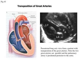
AORTIC ARCHES DEVELOPMENT OF ARTERIES. Dr. Mujahid Khan. Aortic Arches. As the pharyngeal arches form during the 4 th and 5 th weeks, they are supplied by arteries called aortic arches from the aortic sac Initially, the paired dorsal aorta run through the entire length of the embryo

E N D
AORTIC ARCHESDEVELOPMENT OF ARTERIES Dr. Mujahid Khan
Aortic Arches • As the pharyngeal arches form during the 4th and 5th weeks, they are supplied by arteries called aortic arches from the aortic sac • Initially, the paired dorsal aorta run through the entire length of the embryo • They soon fuse to form a single dorsal aorta, just caudal to pharyngeal arches
Aortic Arches • The aortic arches arise from the aortic sac and terminate in the dorsal aorta of the ipsilateral side • Though six pairs of aortic arches usually develop • All are not present at the same time • By the time the sixth pair of aortic arches has formed, the first two pairs disappear • During the eighth week, the aortic arch pattern is transformed to final fetal arterial arrangement
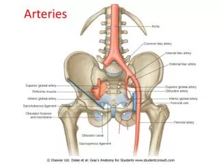
Intersegmental Arteries • Thirty or so branches of the dorsal aorta pass between and carry blood to the somites and their derivatives, called intersegmental arteries • The dorsal intersegmental arteries in the neck join to form a longitudinal artery on each side, the vertebral artery • In the thorax, the dorsal intersegmental arteries persist as intercostal arteries
Intersegmental Arteries • Most of the dorsal intersegmental arteries in the abdomen become lumbar arteries • The 5th pair of lumbar intersegmental arteries remains as the common iliac arteries • In the sacral region, the intersegmental arteries form the lateral sacral arteries • The caudal end of dorsal aorta becomes the median sacral artery
Derivatives of 1st pair • These arteries largely disappear but the remaining parts form the maxillary arteries • Supply the ears, teeth, and muscles of the eye and face • May also contribute to the formation of the external carotid arteries
Derivatives of 2nd pair • Dorsal part of these arteries persist and form the stem of the stapedial arteries • These are small vessels that run through the ring of the stapes • Stapes is a small bone in the ear
Derivatives of 3rd pair • Proximal parts of these arteries form the common carotid arteries • They supply the structures in the head • Distal part of the third pair of aortic arches join with the dorsal aortae to form the internal carotid arteries • They supply to the ears, orbits, brain, and its meninges
Derivatives of 4th pair • The left fourth aortic arch forms part of the arch of aorta • The proximal part of the arch develops from the aortic sac and the distal part is derived from the left dorsal aorta • The right fourth aortic arch becomes the proximal part of the right subclavian artery
Derivatives of 4th pair • The distal part of the subclavian artery forms from the right dorsal aorta and right seventh intersegmental artery • The left subclavian artery is not derived from an aortic arch, it forms from the left seventh intersegmental artery • Left subclavian artery comes close to the origin of left common carotid artery as development proceeds
Fate of 5th pair • In about 50% embryos the fifth pair of aortic arches are rudimentary vessels, that soon degenerate • Leave no vascular derivatives • In other 50% of embryos, these arteries do not develop
Derivatives of 6th pair Left sixth aortic arch • The proximal part of the arch persists as the proximal part of the left pulmonary artery • The distal part of the arch passes from the left pulmonary artery to the dorsal aorta to form an arterial shunt, the ductus arteriosus
Derivatives of 6th pair Right sixth aortic arch: • The proximal part of the arch persists as the proximal part of the right pulmonary artery • The distal part of the arch degenerates
Aortic Arch Anomalies • Due to many changes involved in transformation of the embryonic pharyngeal arch system of arteries into the adult arterial pattern • Most irregularities result from the persistence of parts of aortic arches that usually disappear, or from disappearance of parts that normally persists
Aortic Arch Anomalies • Coarctation of aorta or constriction of aorta • Double aortic arch • Right arch of aorta
Fetal Circulation • Fetal cardiovascular system is designed to serve prenatal needs • It permits modifications at birth and establish a neonatal circulatory pattern • Prenatally the lungs do not provide gas exchange and pulmonary vessels are vasoconstricted
Transitional Neonatal Circulation • At birth the circulation of fetal blood through the placenta ceases • Infant’s lung expand and begin to function • Three shunts that permitted blood to bypass the liver and lungs close and cease to function • The oval foramen, ductus arteriosus, ductus venosus and umbilical vessels are not needed after birth
Transitional Neonatal Circulation • The sphincter in the ductus venosus constricts • All the blood entering the liver passes through the hepatic sinusoids • Occlusion of the placental circulation causes an immediate fall of blood pressure in the IVC and right atrium
Transitional Neonatal Circulation • Aeration of the lungs at birth is associated with: • Dramatic fall in pulmonary vascular resistance • Marked increase in pulmonary blood flow • Progressive thinning of the walls of pulmonary arteries due to stretching
Adult Derivatives of Fetal Vascular Structures • Intra-abdominal part of the umbilical vein eventually becomes round ligament of liver • Ductus venosus becomes the ligamentum venosum • It passes through the liver from the left branch of the portal vein to the IVC to which it is attached
Adult Derivatives of Fetal Vascular Structures • Intra-abdominal part of umbilical arteries become the medial umbilical ligaments • Proximal parts of these vessels persist as the superior vesical arteries • Functional closure of ductus arteriosus is completed within few days after birth • Ductus arteriosus is converted into ligamentum arteriosum