Download

1 / 10
100 likes | 173 Views
Dissection Guide to the Rat. The Norway rat (Rattus norvegicus) Family:Muridae. Anatomical Terms. Cranial -toward the head Caudal -toward the tail Dorsal -toward the backbone Ventral -toward the belly side Lateral -toward the side Medial -toward the middle

E N D
Dissection Guide to the Rat The Norway rat (Rattus norvegicus) Family:Muridae
Anatomical Terms • Cranial-toward the head • Caudal-toward the tail • Dorsal-toward the backbone • Ventral-toward the belly side • Lateral-toward the side • Medial-toward the middle • Proximal-closer to the the base of a structure • Distal-farther away from a structure
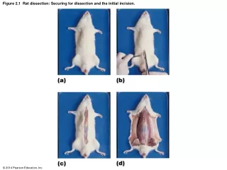
Skinning the Rat • Lay rat on its back • Using a blunt probe, separate the skin from the muscle • Use scissors to cut along ventral midline • Be careful to cut only the skin • Make four lateral incisions • Remove the skin from the body
Muscles to Locate Locate the starred muscles. Draw and label them on your lab report.
Beginning the Muscle Identification The Triceps Brachii
The Internal Anatomy The first incisions were made down the center of the body cavity and down to the feet and through to the arms. These cuts were made so the skin could be neatly folded back and pinned in place.
After the pinning is complete the interior muscle tissues are exposed, then by making careful incisions, the interior organs can be exposed. Carefully cut into the throat being sure not to cut though the masseter, digastric or the mylohyoid. When the body cavity is laid open the first organs that are seen are the large brown livers, cecum (most commonly mistaken for the stomach), and the small and large intestines. After removing the intestinal group the stomach, pancreas, and spleen become much more visible. Moving to the respiratory system that lies under the rib cage, carefully but firmly cut up the center of the ribcage. This is will break the breast bone and the ribs when they are pinned back. Opening the rib cage will expose the heart and lungs
Lab Report • Write your lab report using proper reporting form • Include drawings of the muscles, and internal anatomy • Copy and include a drawing of the skeletal system • Write out your conclusion questions