Download
1 / 25
250 likes | 253 Views
Nephrotic syndrome المتلازمة الكلوية(الكلائية). Lecture-6- Hazem.K.Al-khafaji DM.FICMS University of Al- Qadissiya College of medicine Department of medicine. Nephrotic syndrome الكلائية) ) المتلازمة الكلوية.

E N D
Nephrotic syndromeالمتلازمة الكلوية(الكلائية) • Lecture-6- • Hazem.K.Al-khafaji • DM.FICMS • University of Al-Qadissiya • College of medicine • Department of medicine
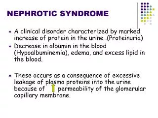
Nephrotic syndromeالكلائية)) المتلازمة الكلوية Definition of Nephrotic Syndrome (NS) : is characterized by heavy proteinuria, hypoproteinemia, generalized oedema and hyperlipidemia. Heavy proteinuria- when urinary protein excretion more than 40mg̸ m2 / hour or 1g/ m2 /24hours. Or ≥3.5gm/ 1.73 m2 /day. Hypoproteinemia- serum albumin < 3g/dl (adult) or 2.5g/dl(children). Hyperlipidemia-serum cholesterol>250mg/dl. Oedema,may be generalized (anasarca). BP may be normal ,decreased or increased. Other consequences are acute or chronic renal failure, hypercoaguable state & depressed immunity.
INTRODUCTION Nephrotic syndrome :Patients present with marked edema, proteinuria, hypoalbuminemia, and often hyperlipidemia .It may be caused by primary (idiopathic) renal disease or by a variety of secondary causes. In adults, diabetes mellitus is the most common secondary cause, and membranous nephropathy is the most common primary causes. In children, minimal change nephropathy is the commonest cause. Venous thrombo-embolism is a possible complication; acute & chronic renal failure and serious bacterial infection are also possible, but much less common. There are no established guidelines on the diagnostic workup or management of nephrotic syndrome. Imaging studies are generally not necessary, and certain blood tests should be used selectively to diagnose specific disorders. Renal biopsy may be useful in some cases to confirm an underlying disease or to identify idiopathic disease . Treatment of most patients should include fluid and sodium restriction, oral or intravenous diuretics, and angiotensin-converting enzyme inhibitors, intravenous albumin not highly recommended. Some adults with nephrotic syndrome may benefit from corticosteroid treatment ,But other immunosuppressive drugs usually required. prophylactic antibiotics, and prophylactic anticoagulation are controversial.& there are certain indications for albumin infusion.
Epidemiology Incidence: About 2 in every 10.000 people experience N.S. Demographics Age Minimal change disease accounts for more than 80% of all cases of nephrotic syndrome in children younger than 10 years but only 10% to 15% of primary cases in adults. Gender Minimal change disease: boys are affected twice as frequently as girls, but the gender ratio is equal in adults.
Pathogenesis Only 150 mg of protein lost in urine daily. The underlying abnormality in nephrotic syndrome is an increase in permeability of the filtering barrier, mainly changes in the podocyte (structure & charge),which leads to massive proteinuria and hypoalbuminemia. The cause of the increased permeability is not well understood.Although,the most common pathology is podocyte injury & changed architecture due to scarring or elements deposition .
PATHOPHYSIOLOGY. lymphocytes, may be responsible for the increase in capillary wall permeability. Alternately, mutations in podocyte proteins (podocin, α-actinin 4) are associated with focal segmental glomerulosclerosis. Steroid-resistant nephrotic syndrome is associated with mutations in NPHS2 (pod In minimal change disease, it is possible that T-cell dysfunction leads to alteration of cytokines, which causes a loss of negatively charged glycoprotein within the glomerular capillary wall. In focal segmental glomerulosclerosis, a plasma factor, perhaps produced by lymphocytes, may be responsible for the increase in capillary wall permeability.
Mechanism of oedema formation is incompletely understood.The following mechanisms are shared : 1-Massive urinary protein loss leads to hypoalbuminemia, which causes a decrease in the plasma oncotic pressure and transudation of fluid from the intravascular compartment to the interstitial space. 2-The depletion of intravascular volume leads to activation of rennin – angiotesin – aldosterone axis resulting in secondary hyperaldosteronism which increase sodium tubular reabsorption Patients may have an “overfilled” or expanded plasma volume in addition to expanded interstitial fluid volume. This may be clinically important if over-rapid diuresis done, it leads to acute renal failure from reduced glomerular blood flow, despite persistent oedema. . 3- primary renal defect leads to retention of sodium and fluid..
What is the cause of hyperlipidemia? HMGCo reductase in liver limits & controls the biosynthesis of cholesterol. Hyperlipidemia is related to the severity of proteinuria and hypoalbuminemia. In NS, hypoalbuminemia causes Up regulation of HMGCo reductase.There is increased production of all lipids and decrease in receptor mediated removal of lipoprotein and lipoprotein lipase. simply it is compensatory mechanism due to increase lipid synthesis & decrease catabolism. Hyperlipidaemia leads to accelerated atherosclerosis & cardiovascular disease.
What is the cause of hypercoaguability ?1- loss of antithrombinIII in the urine.2-altered level & activity of proteins S & C.3-increased hepatic synthesis of fibrinogen.4-Increased platelets aggregation.Complications of hypercoaguability:1- Spontaneous thrombosis ( mostly venous in adult but arterial more common in children).2-Pulmonary embolism.3- Renal vein thrombosis: occur in 30% of patient ,presented as severe flank pain, gross haematuria & abrupt decline in renal function.4- Strokes & myocardial infarctions are also potential complication. The highest incidence in membranous glomerulopathy?
PresentationOedema is the most profound symptom. It started as mild periorbital puffiness that is worsened on lying down, especially at morning ,then pitting oedema of lower exrimeties.Also the patient may had pericardial effusion, pleural effusion & ascites(which developed early in children).subungal oedema may manifest as parallel white lines in the fingernail beds(Leukonychia).In case of severe oedema the patient may generalized swelling or anasarca which is worse in dependent area specially genitalia.
Complications 1-Acute &chronic renal failure 2-Infection: is the major complication of nephrotic syndrome. The increased susceptibility to bacterial infections due to urinary losses of immunoglobulins , defective cell-mediated immunity & other factors contributing are immunosuppressive therapy, malnutrition, and edema specially ascites which act as a potential culture medium. Different types of infection including sepsis, pneumonia, cellulitis, and urinary tract infections. ButSpontaneous bacterial peritonitis is the most frequent type of infection, the commonest organism causing peritonitis is Streptococcus pneumoniae ,but gram-negative bacteria such as Escherichia coli may also be encountered .
3-Thromboembolic events: Both arterial and venous thromboses may be seen, including renal vein thrombosis, pulmonary embolus, sagittal sinus thrombosis, and thrombosis of indwelling arterial and venous catheters.Membranous nephropathy and serum albumin levels less than 2.0 g/dl (20 g/ L) at the highest risk for this complication.. 4- Accelerated atherosclerosis :Leads to increased incidence ofcardiovascular disease such as acute myocardial infarction. 5- Iron resistant hypochromicmicrocytic anemia: due to the urinary loss of iron binding protein(transfferin)
Causes of nephrotic syndrome: Primary nephrotic syndrome: 1-Minimal change N.S :the commonest cause in children less then 10 years old. 2-Membranous nephropathy: the commonest cause in adult. 3-Focal segmental glomerulonephritis. 4-membranoproliferative glomerulonephritis. Secondary N.S: 1-Metabolic;diabetes mellitus,amylodosis. 2- Collagen disease or connective tissue disease :SLE 3- Infection: Hepatitis B, C,HIV, Malaria(P.malariae),Syphilis, Toxoplasmosis.
4-Drugs:Penicillamine,Gold,Non-steroidal anti-inflammatory drugs,Interferon,Mercury,Heroin,Lithium • 5-Immunological & allergens: venom (snake, bee),immunization. • 6- pre-eclampsia. • 7-Genetic disorders: Alport’s syndrome, sickle-cell disease, hereditary glomerulonephritis. 8-Malignancy – Lymphoma,leukemia,solid tumours
- Investigations • Investigations • General Workup • Urinanalysis &24hours urine collection for protein . • Albumin concentration in urine ̸ creatinine concentration in urine should be more than 200mg / mmol in nephrotic syndrome. • Total serum protein , S.albumin &Serum and urine protein electrophoresis. Renal function test as B.urea ,S.creatinine & blood electrolytes. • Lipid profile(Cholesterol,LDL,VLDL,HDL,Trigleyseride). • FBS & HbAc & HbAc1 Renal ultrasound( not mandatory ) • LFT:TSB.SGOT,SGPT,& ALP.Specific serology for selected cases: • HIV, HCV, HBV, ANA, serum cryoglobulins, anti DNA Ab. Genetic studies. • Complement assay: C3,C4.
Indications of renal biopsy : Renal biopsy is often recommended in adult with nephrotic syndrome to establish the pathologic subtype of the disease, to assess disease activity, or to confirm the diagnosis of diseases & also for decision of treatment & finally the prognosis. Children with suspected other causes of N.S rather than minimal change nephropathy i.e if there are haematuria, hypertension, renal insufficiency, hypocomplementaemia, age <1 yr or >8 yr) .
General supportive measures 1- Salt restriction: limit sodium intake to 3 g per day, and may need to restrict fluid intake (to less than approximately 1.5 L per day). 2-Nutritionalsupport:Not recommended to increases protein intake. 3- DIURETICS:Diuretics are the mainstay of medical management; however, there is no evidence to guide drug selection or dosage. diuresis should aim for a target weight loss of (0.5 to 1 kg) per day, to avoid acute renal failure or electrolyte disorders. Loop diuretics, such as furosemide (Lasix) or bumetanide(Burrinex), are most commonly used. Large doses (e.g., 80 to 120 mg of furosemide) are often required and these drugs typically must be given intravenously because of the poor absorption of oral drugs caused by intestinal edema. Low serum albumin levels also limit diuretic effectiveness and necessitate albumin infusion with adjunctive diuretics e.gThiazide diuretics, potassium-sparing diuretics as aldosterone antagonist. these useful as synergistic diuretics. But generally avoid overaggressive diuretic therapy.
4-ACE INHIbitors:Angiotensin-converting enzyme (ACE) inhibitors have been shown to reduce proteinuria and reduce the risk of progression to renal disease in persons with nephrotic syndrome. 5-ALBUMIN:There is no evidence to indicate benefit from treatment with albumin, and the adverse effects, such as hypertension or pulmonary edema, limit its use.Intravenous albumin has been proposed to aid diuresis in massive oedema with poor response to diuretics alone. 6-LIPID-LOWERING TREATMENT:the decision to start lipid-lowering therapy in persons with nephrotic syndrome should be made on the basis as the lipids level. Statins are the mainstay of treatment as theyhave been proven to reduce LDL levels 7-ANTICOAGULATION THERAPYprophylactic anticoagulation recommend to prevent thromboembolism when s.albumin ≤ 2gm/dl ,but nowadays it is controversial
8-Antibiotics: no indications for prophylactic antibiotics or other interventions to prevent infection. Patients who are appropriate candidates should receive pneumococcal vaccination. Treatment of secondary cause in case of secondary N.S. Treatment of primary(idiopathic):CORTICOSTEROIDS: corticosteroids remains controversial in the management of nephrotic syndrome in adults. but it is well established that children respond well to corticosteroid treatment. Classically, minimal change disease responds better to corticosteroids than primary diseases. Immunosuppressive drugs:cyclosporine,Azothioprine,Mycophenolate &others used as1st line in adult & 2nd line in children.
Minimal change glomerulopathy Accounts for 80% of cases of nephrotic syndrome in children younger than 10years, approximately 50% of cases in older children, and 10% to 15% of cases in adults No cause is found in most patients. Disease is immunologically mediated and related to abnormal T-cell function rather than immune-complex deposition Patients usually present with full-blown pure nephrotic syndrome Hypertension and microscopic hematuria are uncommon, but one of these may occur in 20% to 30% of patientsRenal impairment and acute kidney injury can occur but are rare Severe edema, ascites, and pleural effusions may occur Spontaneous remission may occur in rare cases, but it is seldom justified to leave the patient untreated, because persistent nephrotic syndrome causes significant morbidity and mortality The risk of relapse decreases with time, and relapse is often precipitated by a minor upper respiratory tract infection,allergens as foods or bee sting & NSAIDs.
Membranous nephropathy: Most common pathologic entity associated with idiopathic nephrotic syndrome in adults May be primary or secondary to a wide range of diseases. Its pathogenesis is unknown, and treatment is controversial 80% of patients present with nephrotic syndrome, and the remainder present with lesser degrees of proteinuria. Occasionally, patients may present with acute kidney injury as a complication of nephrotic syndrome(30%) Up to 50% of patients are hypertensive at presentation. Tratment with corticosteroids or otheimmunosupressive drugs as cyclosporine A, mycophenolylatemofetil & others.
Choose the most appropriate answer • A 30 years old male patient presented to the emergency unit complaining of dyspnoea for last 7 days. • O̸E:puffy face & both legs looks swelling, but left one with pitting oedema & right one looks more swelling, tense, hot & tender. • Chest examination :bilateral dullness& absence of breathing sound in the lower zones . • Investigations revealed 24 hours urine protein 7gm..S.albumin 1.8gm / l.. • The most likely diagnosis is : • A- Minimal change nephropathy. • B- Goodpasture’s disease. • C- Wegner granulomatosis. • D- Henoch-SchÖnlein purpura • E- Membranous nephropathy.